

| Journal of Clinical Medicine Research, ISSN 1918-3003 print, 1918-3011 online, Open Access |
| Article copyright, the authors; Journal compilation copyright, J Clin Med Res and Elmer Press Inc |
| Journal website http://www.jocmr.org |
Original Article
Volume 10, Number 12, December 2018, pages 928-935
Clinical Impact of Hemorheology on Subclinical Myocardial Injury in Patients with Hypertension
Takashi Hitsumotoa, b
aHitsumoto Medical Clinic, Yamaguchi, Japan
bCorresponding Author: Takashi Hitsumoto, Hitsumoto Medical Clinic, 2-7-7, Takezakicyou, Shimonoseki-City, Yamaguchi 750-0025, Japan
Manuscript submitted October 5, 2018, accepted October 16, 2018
Short title: Hemorheology and hs-cTnT in Hypertension
doi: https://doi.org/10.14740/jocmr3652
| Abstract | ▴Top |
Background: The blood concentration of high-sensitivity cardiac troponin T (hs-cTnT) is a useful biomarker for myocardial injury or the pathogenesis of hypertension. Little is known about the relationship between hemorheology and myocardial injury in patients with hypertension. This cross-sectional study aimed to clarify the clinical impact of hemorheology on subclinical myocardial injury assessed with a microchannel array flow analyzer (MC-FAN) and its impact on hs-cTnT in patients with hypertension.
Methods: A total of 447 outpatients (men: 181; women: 266; mean age: 65 ± 13 years), with no history of cardiovascular disease, including admission for heart failure, who were undergoing treatment for hypertension, were enrolled. Whole blood passage time (WBPT) as a marker of hemorheology was measured with a MC-FAN, and the relationship between hs-cTnT levels and various clinical parameters, including WBPT, was examined.
Results: hs-cTnT levels were detected in 400 patients (89.5%). WBPT was significantly higher in patients with detectable hs-cTnT levels than in those with undetectable hs-cTnT levels (60.5 ± 16.8 s versus 50.2 ± 14.2 s, P < 0.001). In patients with detectable hs-cTnT levels, there was a significant positive correlation between WBPT and hs-cTnT level (r = 0.33; P < 0.001). Multiple regression analysis revealed that WBPT was an independent variable when hs-cTnT was a subordinate factor (β = 0.15; P < 0.01). Receiver-operating characteristic curve analysis indicated that a cutoff value for WBPT of 55.6 s yielded the largest area under the curve (0.744; P < 0.001) for discriminating high hs-cTnT levels as ≥ 0.014 ng/mL.
Conclusion: The results indicate that WBPT is independently associated with hs-cTnT in hypertensive patients with no history of cardiovascular events, suggesting that impairment of hemorheology in small cardiac vessels causes subclinical myocardial injury. In addition, the study suggests that progression of myocardial injury can be prevented by maintaining WBPT at approximately ≤ 55 s.
Keywords: Hemorheology; Microchannel array flow analyzer; High-sensitivity troponin T; Advanced glycation end products; Cardio-ankle vascular index; Oxidative stress; Hypertension
| Introduction | ▴Top |
Recent clinical studies have demonstrated that the blood concentration of cardiac troponin T (high-sensitivity cardiac troponin T (hs-cTnT)) can be measured by a highly sensitive assay. The blood concentration of hs-cTnT is a useful biomarker to evaluate myocardial injury or to predict cardiovascular events at the clinical stage [1]. In addition, several studies have reported the clinical significance of hs-cTnT in patients with hypertension [2-5].
Impairment of hemorheology is an important risk factor for cardiovascular disease as well as atherosclerosis [6, 7]. In recent years, the microchannel array flow analyzer (MC-FAN), which is a commercial device that assesses hemorheology using microscopic images, has been introduced in clinical settings [8]. MC-FAN has a simple methodology and is superior to other methods in the accuracy of channel dimensions and high reproducibility. Clinical studies have demonstrated relationships between increased whole blood passage time (WBPT), measured with MC-FAN, and cardiovascular risk factors or cardiovascular events [9-12].
Little is known about the relationships between hemorheology and myocardial injury in patients with hypertension. This study aimed to elucidate the association of WBPT with hs-cTnT in patients with hypertension and without apparent cardiovascular disease, including heart failure.
| Patients and Methods | ▴Top |
Study population
This study was conducted at the Hitsumoto Medical Clinic in Shimonoseki City, Japan, between March 2016 and February 2018. The study population comprised 447 outpatients (males: 181 (40.5%) and females: 266 (59.5%); mean age: 65 ± 13 years) undergoing treatment for hypertension. No patient had a history of cardiovascular events, such as coronary artery, cerebrovascular, or perivascular disease or admission for heart failure. All patients provided informed consent, and the study protocol was approved by the Local Ethics Committee of the Hitsumoto Medical Clinic (approval number: 2016-02).
Evaluation of hemorheology by MC-FAN
The evaluation of hemorheology was performed by measuring WBPT with an MC-FAN HR300 rheometer (MC Healthcare, Tokyo, Japan), as previously reported (Fig. 1) [8, 10]. Briefly, the microchannel passage time for 100 µL of physiological saline as a control was measured, followed by the microchannel passage time for 100 µL of a heparinized whole blood sample obtained from a patient. The WBPT for patients was corrected for the passage time of physiological saline. The microchannel formation was 7-µm wide, 30-µm long and 4.5-µm deep. WBPT was measured within 60 min of blood sampling. The inter- and intra-assay coefficients of variation for WBPT were 8% and 5%, respectively.
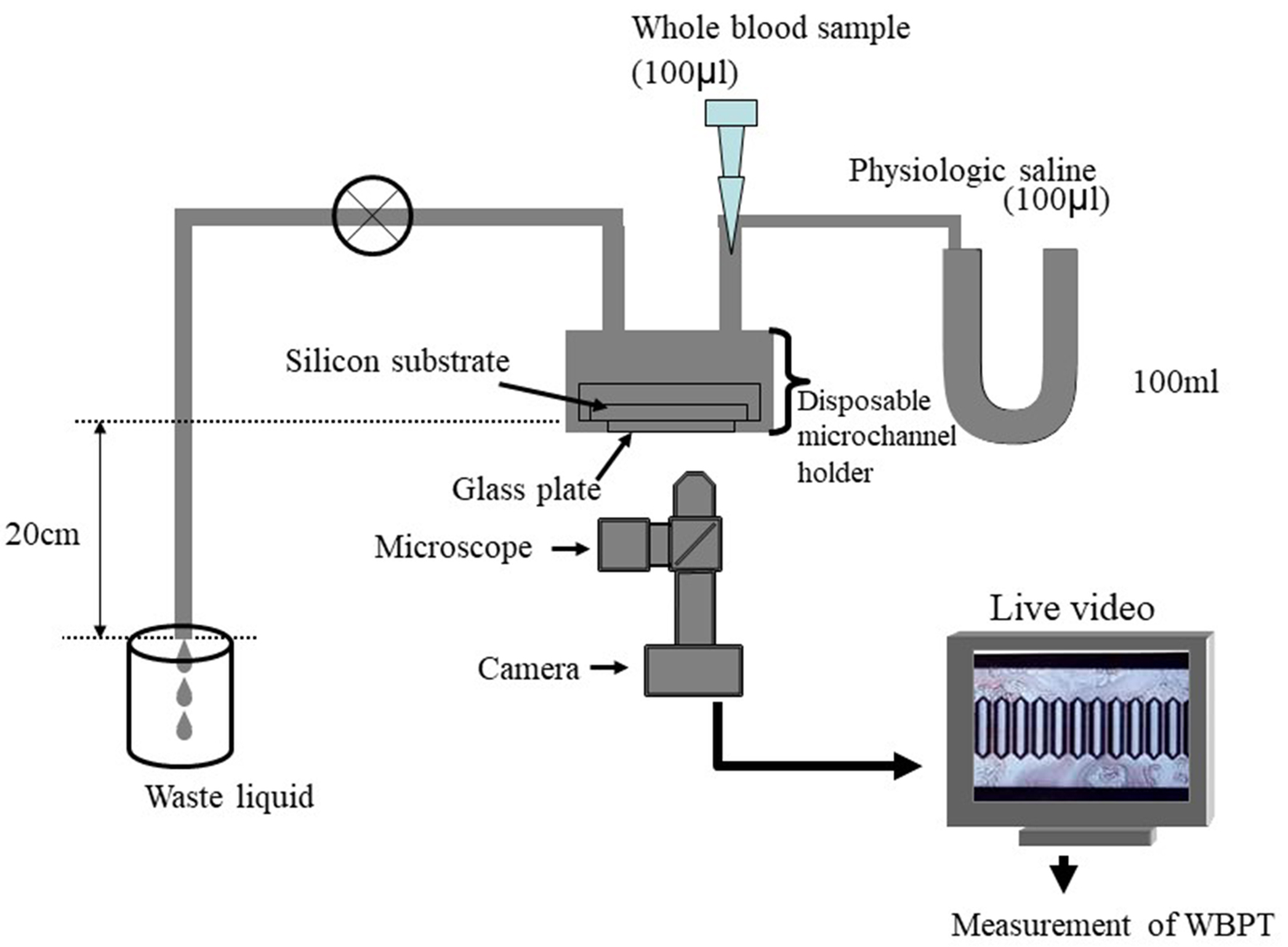 Click for large image | Figure 1. System of microchannel array flow analyzer. The microchannel passage time of 100 µL of physiological saline was measured as a control, and then that of venous whole blood obtained from the subjects with 5% heparinization was determined. The WBPT of the subjects was expressed after correction for the passage time of physiological saline. Inter- and intra-assay coefficients of variation for WBPT were 8% and 5%, respectively. WBPT: whole blood passage time. |
Estimation of clinical parameters
Various clinical parameters, classic coronary risk factors, exercise habits, glucose-related parameters, blood cell count, kidney function, left ventricular hypertrophy estimated by electrocardiography, brain natriuretic peptide (BNP), oxidative stress, arterial function and hs-cTnT levels were evaluated. The degree of obesity was evaluated by the body mass index, calculated as the weight in kilograms divided by the height in square meters. Exercise habits were considered positive for those who performed aerobic exercise for more than 30 min three times a week. Current smoking was defined as smoking at least one cigarette per day during the previous 28 days. Treatment with antihypertensive drugs was stopped 24 h or more before measurement, and right brachial blood pressure was measured twice with a mercurial sphygmomanometer with the patient in a sitting position. The average of two readings was used to determine systolic and diastolic blood pressure. Dyslipidemia was defined as low-density lipoprotein cholesterol level ≥ 140 mg/dL, high-density lipoprotein cholesterol level ≤ 40 mg/dL, triglyceride level ≥ 150 mg/dL or the use of antidyslipidemic medication. Skin autofluorescence (AF) as a marker of advanced glycation end products (AGEs) in the tissues was measured with a commercial instrument (AGE ReaderTM, DiagnOptics, Groningen, The Netherlands), as described previously [13]. Briefly, AF was defined as the average light intensity per nanometer in the range between 300 and 420 nm. AF levels were expressed in arbitrary units. All measurements were taken at the volar side of the lower arm, approximately 10 - 15 cm below the elbow fold, with the patient in a seated position. The severity of left ventricular hypertrophy was evaluated using Cornell (R wave in aVL + S wave in V3) electrocardiographic voltage calculations [14]. Arterial function was evaluated by the cardio-ankle vascular index (CAVI) using a VaSera CAVI instrument (Fukuda Denshi, Tokyo, Japan) according to previously described methods [15]. Briefly, brachial and ankle pulse waves were determined with inflatable cuffs by maintaining the pressure between 30 and 50 mm Hg to ensure a minimal effect on systemic hemodynamics from the pressure of the cuffs. Blood pressure and pulse pressure were simultaneously measured with the patient in the supine position. CAVI was measured after the patient rested for 10 min in a quiet room. CAVI was calculated by the formula CAVI = a((2ρ/ΔP) × ln(Ps/Pd)PWV2) + b, where a and b are constants, ρ is blood density, ΔP is Ps - Pd, Ps is systolic blood pressure, Pd is diastolic blood pressure and PWV is pulse wave velocity.
Evaluation of blood parameters
Blood samples were collected from the antecubital vein in the morning after 12 h of fasting. Total cholesterol and triglyceride concentrations were measured by standard enzymatic methods. Serum high-density lipoprotein cholesterol concentrations were measured by selective inhibition, and serum low-density lipoprotein cholesterol concentrations were measured by the Friedewald equation [16]. Patients with a serum triglyceride concentration ≥ 400 mg/dL were excluded, considering the accuracy of this method. Glucose and insulin concentrations were measured by the glucose oxidase method and enzyme immunoassay, respectively. To measure insulin resistance, homeostatic model assessment of insulin resistance (HOMA-IR) was calculated as follows [17]: HOMA-IR = fasting glucose concentration (mg/dL) × fasting insulin concentration (µg/mL)/405. Estimated glomerular filtration rate (eGFR) was calculated by the adjusted Modification of Diet in Renal Disease Study equation, which was proposed by the working group of the Japanese Chronic Kidney Disease Initiative [18]. The blood concentration of BNP was measured with a commercial kit (SHIONOSPOT Reader; Shionogi & Co., Osaka, Japan). The derivatives of reactive oxygen metabolites (d-ROMs) test as a marker of oxidative stress in vivo was conducted with a commercial kit (Diacron, Grosseto, Italy) [19]. hs-cTnT levels were also measured with a commercial kit (Roche Diagnostics, Basel, Switzerland) [20]. In the hs-cTnT assay, the lower limit of detection was 0.003 ng/mL.
Statistical analysis
The data were analyzed by Stat View-J 5.0 (HULINKS, Tokyo, Japan) and MedCalc for Windows version 14.8.1 (MedCalc Software, Ostend, Belgium). The data are expressed as means ± SD. Between-group comparisons were performed by Student’s t-test or the Mann-Whitney U test, and the correlation coefficient was estimated by Spearman rank-order correlation analysis. Multivariate analysis was conducted by multiple regression analysis. Receiver-operating characteristic (ROC) curves were constructed, and the Youden index was used to determine the optimal cutoff for WBPT for determining high hs-cTnT levels. A P value < 0.05 was considered to indicate statistical significance.
| Results | ▴Top |
Table 1 presents the patients’ characteristics. hs-cTnT levels were detected in 400 patients (89.5%). Age, systolic blood pressure, presence of diabetes mellitus, fasting blood glucose level, HOMA-IR, skin AF, Cornell voltage, BNP, d-ROMs test, CAVI and frequency of antidiabetic medication were significantly higher and eGFR was significantly lower in patients with detectable hs-cTnT levels. WBPT was significantly higher in patients with detectable hs-cTnT levels. Correlations among hs-cTnT level, WBPT and various clinical parameters in patients with detectable hs-cTnT levels are presented in Table 2. Gender, age, presence of diabetes mellitus, fasting blood glucose level, skin AF, eGFR, Cornell voltage, BNP, d-ROMs test and CAVI were significantly correlated with hs-cTnT levels. Body mass index, exercise habits, current smoking status, presence of diabetes mellitus, skin AF, white blood cell count, hematocrit, eGFR, d-ROMs test and CAVI were significantly correlated with WBPT. The correlations between hs-cTnT level and WBPT are shown in Figure 2. Significantly positive correlations were observed between the two parameters. Table 3 shows the results of multiple regression analysis for hs-cTnT levels or WBPT as a subordinate factor. Explanatory factors were selected either by checking multicollinearity among variables or by the stepwise method. Skin AF, CAVI, WBPT, age, d-ROMs test, eGFR and BNP level were selected as independent variables when hs-cTnT level was used as a subordinate factor; CAVI, hs-cTnT level, d-ROMs test, skin AF, hematocrit, exercise habits and current smoking status were selected as independent variables when WBPT was used as a subordinate factor. Figure 3 shows the ROC curve analysis for detection of high hs-cTnT levels as ≥ 0.014 ng/mL based on WBPT. A WBPT cutoff of 55.6 s yielded the largest area under the curve, 0.744 (95% confidence interval: 0.699 - 0.784; P < 0.001), indicating a sensitivity of 85.7% and a specificity of 57.0% for discriminating hs-cTnT levels as ≥ 0.014 ng/mL.
 Click to view | Table 1. Patient Characteristics |
Click to view | Table 2. Correlation Between hs-cTnT, WBPT and Clinical Parameters in hs-cTnT Detectable Patients |
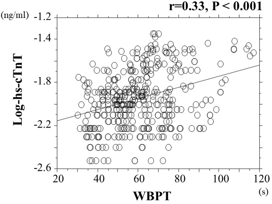 Click for large image | Figure 2. Relationship between hs-cTnT and WBPT. There is significantly positive correlation between log-hs-cTnT and WBPT (r = 0.33, P < 0.001). hs-cTnT: high-sensitivity troponin T; WBPT: whole blood passage time. |
Click to view | Table 3. Multiple Regression Analysis |
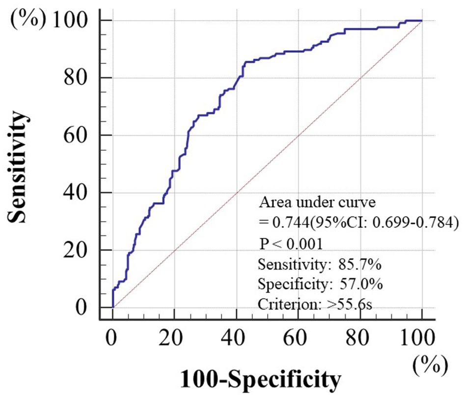 Click for large image | Figure 3. Cutoff value of WBPT for discriminating hs-cTnT levels as ≥ 0.014 ng/mL. Receiver-operating characteristic indicated that a WBPT cutoff of 55.6 s yielded the largest area under the curve, 0.744 (95% confidence interval: 0.699 - 0.784; P < 0.001), indicating a sensitivity of 85.7% and a specificity of 57.0% for discriminating hs-cTnT levels as ≥ 0.014 ng/mL. WBPT: whole blood passage time; hs-cTnT: high-sensitivity troponin T. |
| Discussion | ▴Top |
Yagi et al reported that the mean WBPT in healthy controls with no history of traditional cardiovascular risk factors, such as hypertension, hypercholesterolemia and diabetes mellitus, was 37.1 s [21], whereas the mean WBPT in the present study was 59.4 s. Thus, these results indicate that the impairment of hemorheology in hypertensive patients was progressive compared with that in healthy controls. Furthermore, independent associations between WBPT and hs-cTnT levels in this study suggest that the impairment of hemorheology affects the progression of subclinical myocardial injury in patients with hypertension. Skin AF, CAVI and d-ROMs test were selected as independent variables for both hs-cTnT and WBPT as subordinate factors.
Hemorheology assessed by MC-FAN is an in vitro measurement that uses artificial blood vessels, with a 7-µm wide, 30-µm long and 4.5-µm deep vessel lumen. Thus, assessment of hemorheology by MC-FAN is assumed to correspond to values obtained for small vessels. Small vessels are considered to be present in a portion of skin biopsies along with systemic vessels, including heart vessels. On the contrary, the results of this study indicated that skin AF as a marker of AGEs in tissues was selected as an independent variable when hs-cTnT was a subordinate factor. In addition, skin AF had an independent association with WBPT. Skin AF is reported to reflect pentosidine levels, which are major components of AGEs on the volar side of the lower arm according to the results of skin biopsy [22]. Therefore, impairment of hemorheology is a possible cause of the accumulation of AGEs in systemic tissues, including the heart. In addition, Hofmann et al have clarified a significant relationship between AGE-modified cardiac tissue collagen and skin AF [23]. Several pathways by which AGEs or their receptors influence myocardial injury have been reported [24-26]. Furthermore, basic studies have indicated that AGEs influence hemorheology by mechanisms such as platelet aggregation, leukocyte-endothelial interaction and morphological changes in the erythrocyte membrane [27-29]. Thus, the results of this and previous studies indicate that hemorheology and AGEs are associated not only on the volar side of the lower arm but also in small cardiac vessels, consequently leading to myocardial injury in patients with hypertension.
CAVI is explored as a marker of arterial stiffness, which is independently associated with blood pressure [15]. Several recent studies have reported the significance of left ventricular dysfunction in the progression of myocardial injury. Furthermore, increases in aortic artery stiffness or arterial reflection wave are known to be caused by left ventricular dysfunction [30, 31]. Therefore, the independent relationship between CAVI and hs-cTnT identified in this study is thought to reflect subclinical myocardial injury via left ventricular dysfunction resulting from an increase in afterload or arterial reflection wave. In addition, some researchers have reported that CAVI reflects endothelial function [32, 33]. Endothelial dysfunction has also been known to cause left ventricular dysfunction and myocardial injury [34, 35]. Thus, the results of this study indicate the importance of arterial dysfunction in subclinical myocardial injury in patients with hypertension. CAVI was also selected as an independent variable for WBPT as a subordinate factor in multivariate analysis. Some studies have reported relationships between WBPT and arterial function, such as arterial stiffness, endothelial function and vascular resistance [9, 21, 36, 37]. Therefore, increased WBPT due to increased hs-cTnT levels may be partly explained by arterial dysfunction.
A number of studies indicate that oxidative stress has a crucial role in the pathogenesis of hypertension [38, 39]. The results of the present study also indicate that the d-ROMs test, as a marker of oxidative stress, is associated with both myocardial injury and hemorheology in patients with hypertension. Several pathways have been identified by which oxidative stress causes myocardial injury, such as dysfunction of the mitochondrial electron transport complex, activity of nicotinamide adenine dinucleotide phosphate oxidase, apoptosis of myocardial cells and nitric oxide synthase uncoupling [40-42]. In addition, several mechanisms are also reported by which oxidative stress causes impairment of hemorheology, such as platelet aggregation and elevation of plasma viscosity [43, 44]. Thus, oxidative stress is an important target factor for the prevention of both the progression of myocardial injury and impairment of hemorheology in patients with hypertension.
It is useful to have target cutoff levels of WBPT for predicting abnormal hs-cTnT levels in clinical settings. This study clarifies the clinical usefulness of assessing WBPT to detect hs-cTnT levels as high as ≥ 0.014 ng/mL, which is shown to be discriminatory of increased hs-cTnT levels [45]. ROC curve analysis for hs-cTnT levels as high as ≥ 0.014 ng/mL indicated that a cutoff value for WBPT of 55.6 s yielded the largest area under the curve (0.744) for discriminating high hs-cTnT levels. Although this was a cross-sectional study, we believe that it is possible to evaluate the risk of the progression of myocardial injury in patients with hypertension by measuring WBPT. Some clinical studies have indicated that medication, exercise habits and smoking cessation improve hemorheology [46-51]. In this cross-sectional study, exercise habits and current smoking status were independently associated with WBPT, although no significant association was observed between medication and WBPT. We suggest that progression of myocardial injury can be prevented by interventions, such as lifestyle modification or aggressive use of medication, which is effective to improve hemorheology in hypertensive patients to maintain WBPT at approximately ≤ 55 s.
This study has several limitations. First, the contents of medical treatment may have influenced the study results. Second, angiography, computed tomography, magnetic resonance imaging and echocardiography were not performed; thus, asymptomatic cardiovascular diseases may have remained undetected. Third, assessment of hemorheology by MC-FAN is an in vitro method that uses artificial blood vessels; therefore, the hemorheological data obtained might differ from those obtained in vivo because of the influence of vascular factors, such as endothelial or smooth muscle cells. However, the results of this study indicate that WBPT is a considerable risk factor for the progression of subclinical myocardial injury in patients with hypertension. Finally, this study was cross-sectional and conducted in a single unit with a relatively small sample size. Future multicenter prospective studies, including intervention therapies, will be required to confirm the results of this study.
Conclusions
WBPT is independently associated with hs-cTnT in hypertensive patients with no history of cardiovascular events, suggesting that impairment of hemorheology in small cardiac vessels causes subclinical myocardial injury. In addition, this study suggests that the progression of myocardial injury can be prevented by maintaining WBPT at approximately ≤ 55 s. Additional prospective studies, including intervention therapies, will be required to confirm the results of this study.
Conflict of Interest
The author has no competing interests.
Grant Support
None.
Financial Disclosure
The author has reported no conflicts of interest.
| References | ▴Top |
- Twerenbold R, Jaffe A, Reichlin T, Reiter M, Mueller C. High-sensitive troponin T measurements: what do we gain and what are the challenges? Eur Heart J. 2012;33(5):579-586.
doi pubmed - Sato Y, Yamamoto E, Sawa T, Toda K, Hara T, Iwasaki T, Fujiwara H, et al. High-sensitivity cardiac troponin T in essential hypertension. J Cardiol. 2011;58(3):226-231.
doi pubmed - Miao DM, Zhang LP, Yu HP, Zhang JY, Xiao WK, Ye P. Serum levels of high-sensitivity troponin T: a novel marker for left ventricular remodeling and performance in hypertensive subjects. Genet Mol Res. 2014;13(3):5143-5153.
doi pubmed - Cayli M, Gur M, Elbasan Z, Seker T, Turkoglu C, Kaypakli O, Sahin DY, et al. High-sensitivity cardiac troponin T predicts nondipper hypertension in newly diagnosed hypertensive patients. J Clin Hypertens (Greenwich). 2013;15(10):731-736.
- Hitsumoto T. Arterial Velocity Pulse Index as a Novel Marker of Atherosclerosis Using Pulse Wave Analysis on High Sensitivity Troponin T in Hypertensive Patients. Cardiol Res. 2017;8(2):36-43.
doi pubmed - Tzoulaki I, Murray GD, Lee AJ, Rumley A, Lowe GD, Fowkes FG. Relative value of inflammatory, hemostatic, and rheological factors for incident myocardial infarction and stroke: the Edinburgh Artery Study. Circulation. 2007;115(16):2119-2127.
doi pubmed - Cowan AQ, Cho DJ, Rosenson RS. Importance of blood rheology in the pathophysiology of atherothrombosis. Cardiovasc Drugs Ther. 2012;26(4):339-348.
doi pubmed - Kikuchi Y, Sato K, Mizuguchi Y. Modified cell-flow microchannels in a single-crystal silicon substrate and flow behavior of blood cells. Microvasc Res. 1994;47(1):126-139.
doi pubmed - Satoh N, Kotani K, Wada H, Himeno A, Shimada S, Sasaki Y, Yamada K, et al. Unfavorable blood rheology is closely associated with arterial stiffness in obese patients. Endocr J. 2009;56(7):915-918.
doi pubmed - Hitsumoto T. Factors affecting impairment of blood rheology in obese subjects. J Cardiol. 2012;60(5):401-406.
doi pubmed - Yoshida K, Kimura T, Aoki T, Tsunekawa K, Araki O, Shoho Y, Nara M, et al. Fasting serum insulin levels and insulin resistance are associated with blood rheology in Japanese young adults without diabetes. J Int Med Res. 2016;44(3):496-507.
doi pubmed - Hitsumoto T. Usefulness of the Whole Blood Passage Time as a Predictor of Primary Cardiovascular Events in Patients With Traditional Cardiovascular Risk Factors. Cardiol Res. 2018;9(4):231-238.
doi pubmed - Meerwaldt R, Links TP, Graaff R, Hoogenberg K, Lefrandt JD, Baynes JW, Gans RO, et al. Increased accumulation of skin advanced glycation end-products precedes and correlates with clinical manifestation of diabetic neuropathy. Diabetologia. 2005;48(8):1637-1644.
doi pubmed - Okin PM, Devereux RB, Liu JE, Oikarinen L, Jern S, Kjeldsen SE, Julius S, et al. Regression of electrocardiographic left ventricular hypertrophy predicts regression of echocardiographic left ventricular mass: the LIFE study. J Hum Hypertens. 2004;18(6):403-409.
doi pubmed - Shirai K, Utino J, Otsuka K, Takata M. A novel blood pressure-independent arterial wall stiffness parameter; cardio-ankle vascular index (CAVI). J Atheroscler Thromb. 2006;13(2):101-107.
doi pubmed - Friedewald WT, Levy RI, Fredrickson DS. Estimation of the concentration of low-density lipoprotein cholesterol in plasma, without use of the preparative ultracentrifuge. Clin Chem. 1972;18(6):499-502.
pubmed - Matthews DR, Hosker JP, Rudenski AS, Naylor BA, Treacher DF, Turner RC. Homeostasis model assessment: insulin resistance and beta-cell function from fasting plasma glucose and insulin concentrations in man. Diabetologia. 1985;28(7):412-419.
doi pubmed - Imai E, Horio M, Nitta K, Yamagata K, Iseki K, Hara S, Ura N, et al. Estimation of glomerular filtration rate by the MDRD study equation modified for Japanese patients with chronic kidney disease. Clin Exp Nephrol. 2007;11(1):41-50.
doi pubmed - Cesarone MR, Belcaro G, Carratelli M, Cornelli U, De Sanctis MT, Incandela L, Barsotti A, et al. A simple test to monitor oxidative stress. Int Angiol. 1999;18(2):127-130.
pubmed - Mingels A, Jacobs L, Michielsen E, Swaanenburg J, Wodzig W, van Dieijen-Visser M. Reference population and marathon runner sera assessed by highly sensitive cardiac troponin T and commercial cardiac troponin T and I assays. Clin Chem. 2009;55(1):101-108.
doi pubmed - Yagi H, Sumino H, Aoki T, Tsunekawa K, Araki O, Kimura T, Nara M, et al. Impaired blood rheology is associated with endothelial dysfunction in patients with coronary risk factors. Clin Hemorheol Microcirc. 2016;62(2):139-150.
doi pubmed - Meerwaldt R, Graaff R, Oomen PHN, Links TP, Jager JJ, Alderson NL, Thorpe SR, et al. Simple non-invasive assessment of advanced glycation endproduct accumulation. Diabetologia. 2004;47(7):1324-1330.
doi pubmed - Hofmann B, Jacobs K, Navarrete Santos A, Wienke A, Silber RE, Simm A. Relationship between cardiac tissue glycation and skin autofluorescence in patients with coronary artery disease. Diabetes Metab. 2015;41(5):410-415.
doi pubmed - Ma H, Li SY, Xu P, Babcock SA, Dolence EK, Brownlee M, Li J, et al. Advanced glycation endproduct (AGE) accumulation and AGE receptor (RAGE) up-regulation contribute to the onset of diabetic cardiomyopathy. J Cell Mol Med. 2009;13(8B):1751-1764.
doi pubmed - Brett J, Schmidt AM, Yan SD, Zou YS, Weidman E, Pinsky D, Nowygrod R, et al. Survey of the distribution of a newly characterized receptor for advanced glycation end products in tissues. Am J Pathol. 1993;143(6):1699-1712.
pubmed - Yu Y, Wang L, Delguste F, Durand A, Guilbaud A, Rousselin C, Schmidt AM, et al. Advanced glycation end products receptor RAGE controls myocardial dysfunction and oxidative stress in high-fat fed mice by sustaining mitochondrial dynamics and autophagy-lysosome pathway. Free Radic Biol Med. 2017;112:397-410.
doi pubmed - Hasegawa Y, Suehiro A, Higasa S, Namba M, Kakishita E. Enhancing effect of advanced glycation end products on serotonin-induced platelet aggregation in patients with diabetes mellitus. Thromb Res. 2002;107(6):319-323.
doi - Morigi M, Angioletti S, Imberti B, Donadelli R, Micheletti G, Figliuzzi M, Remuzzi A, et al. Leukocyte-endothelial interaction is augmented by high glucose concentrations and hyperglycemia in a NF-kB-dependent fashion. J Clin Invest. 1998;101(9):1905-1915.
doi pubmed - Awasthi S, Gayathiri SK, Ramya R, Duraichelvan R, Dhason A, Saraswathi NT. Advanced Glycation-Modified Human Serum Albumin Evokes Alterations in Membrane and Eryptosis in Erythrocytes. Appl Biochem Biotechnol. 2015;177(5):1013-1024.
doi pubmed - Jaroch J, Loboz Grudzien K, Bociaga Z, Kowalska A, Kruszynska E, Wilczynska M, Dudek K. The relationship of carotid arterial stiffness to left ventricular diastolic dysfunction in untreated hypertension. Kardiol Pol. 2012;70(3):223-231.
pubmed - Higashi H, Okayama H, Saito M, Morioka H, Aono J, Yoshii T, Hiasa G, et al. Relationship between augmentation index and left ventricular diastolic function in healthy women and men. Am J Hypertens. 2013;26(11):1280-1286.
doi pubmed - Endo K, Saiki A, Ohira M, Miyashita Y, Shirai K. Cardio-ankle vascular index may reflect endothelial function in type 2 diabetes. Int J Clin Pract. 2011;65(11):1200-1201.
doi pubmed - Kim KJ, Lee BW, Kim HM, Shin JY, Kang ES, Cha BS, Lee EJ, et al. Associations between cardio-ankle vascular index and microvascular complications in type 2 diabetes mellitus patients. J Atheroscler Thromb. 2011;18(4):328-336.
doi pubmed - Zizek B, Poredos P. Increased left ventricular mass and diastolic dysfunction are associated with endothelial dysfunction in normotensive offspring of subjects with essential hypertension. Blood Press. 2007;16(1):36-44.
doi pubmed - Marti V, Aymat R, Ballester M, Garcia J, Carrio I, Auge JM. Coronary endothelial dysfunction and myocardial cell damage in chronic stable idiopathic dilated cardiomyopathy. Int J Cardiol. 2002;82(3):237-245.
doi - Hitsumoto T. Impact of hemorheology assessed by the microchannel method on pulsatility index of the common carotid artery in patients with type 2 diabetes mellitus. J Clin Med Res. 2017;9(7):579-585.
doi pubmed - Hitsumoto T. Relationship between hemorheology assessed using microchannel array flow analyzer and kidney function in hypertensive patients. Cardiol Res. 2017;8(4):147-153.
doi pubmed - Sinha N, Dabla PK. Oxidative stress and antioxidants in hypertension-a current review. Curr Hypertens Rev. 2015;11(2):132-142.
doi pubmed - Montezano AC, Dulak-Lis M, Tsiropoulou S, Harvey A, Briones AM, Touyz RM. Oxidative stress and human hypertension: vascular mechanisms, biomarkers, and novel therapies. Can J Cardiol. 2015;31(5):631-641.
doi pubmed - Li SY, Yang X, Ceylan-Isik AF, Du M, Sreejayan N, Ren J. Cardiac contractile dysfunction in Lep/Lep obesity is accompanied by NADPH oxidase activation, oxidative modification of sarco(endo)plasmic reticulum Ca2+-ATPase and myosin heavy chain isozyme switch. Diabetologia. 2006;49(6):1434-1446.
doi pubmed - Boudina S, Abel ED. Diabetic cardiomyopathy, causes and effects. Rev Endocr Metab Disord. 2010;11(1):31-39.
doi pubmed - Ansley DM, Wang B. Oxidative stress and myocardial injury in the diabetic heart. J Pathol. 2013;229(2):232-241.
doi pubmed - Tzoulaki I, Murray GD, Lee AJ, Rumley A, Lowe GD, Fowkes FG. Inflammatory, haemostatic, and rheological markers for incident peripheral arterial disease: Edinburgh Artery Study. Eur Heart J. 2007;28(3):354-362.
doi pubmed - Podrez EA, Byzova TV, Febbraio M, Salomon RG, Ma Y, Valiyaveettil M, Poliakov E, et al. Platelet CD36 links hyperlipidemia, oxidant stress and a prothrombotic phenotype. Nat Med. 2007;13(9):1086-1095.
doi pubmed - Saunders JT, Nambi V, de Lemos JA, Chambless LE, Virani SS, Boerwinkle E, Hoogeveen RC, et al. Cardiac troponin T measured by a highly sensitive assay predicts coronary heart disease, heart failure, and mortality in the Atherosclerosis Risk in Communities Study. Circulation. 2011;123(13):1367-1376.
doi pubmed - Yamada K, Hirayama T, Hasegawa Y. Antiplatelet effect of losartan and telmisartan in patients with ischemic stroke. J Stroke Cerebrovasc Dis. 2007;16(5):225-231.
doi pubmed - Shamanaev AY, Aliev OI, Anishchenko AM, Sidehmenova AV, Plotnikov MB. Hemorheological effects of amlodipine in spontaneously hypertensive rats. Indian J Pharmacol. 2017;49(4):312-316.
doi pubmed - Ott C, Raff U, Schmidt S, Kistner I, Friedrich S, Bramlage P, Harazny JM, et al. Effects of saxagliptin on early microvascular changes in patients with type 2 diabetes. Cardiovasc Diabetol. 2014;13:19.
doi pubmed - Tirnaksiz E, Pamukcu B, Oflaz H, Nisanci Y. Effect of high dose statin therapy on platelet function; statins reduce aspirin-resistant platelet aggregation in patients with coronary heart disease. J Thromb Thrombolysis. 2009;27(1):24-28.
doi pubmed - Coppola L, Grassia A, Coppola A, Tondi G, Peluso G, Mordente S, Gombos G. Effects of a moderate-intensity aerobic program on blood viscosity, platelet aggregation and fibrinolytic balance in young and middle-aged sedentary subjects. Blood Coagul Fibrinolysis. 2004;15(1):31-37.
doi pubmed - Shimada S, Hasegawa K, Wada H, Terashima S, Satoh-Asahara N, Yamakage H, Kitaoka S, et al. High blood viscosity is closely associated with cigarette smoking and markedly reduced by smoking cessation. Circ J. 2011;75(1):185-189.
doi pubmed
This article is distributed under the terms of the Creative Commons Attribution Non-Commercial 4.0 International License, which permits unrestricted non-commercial use, distribution, and reproduction in any medium, provided the original work is properly cited.
Journal of Clinical Medicine Research is published by Elmer Press Inc.