

| Journal of Clinical Medicine Research, ISSN 1918-3003 print, 1918-3011 online, Open Access |
| Article copyright, the authors; Journal compilation copyright, J Clin Med Res and Elmer Press Inc |
| Journal website http://www.jocmr.org |
Case Report
Volume 1, Number 4, October 2009, pages 237-239
A Rare Case of Primary Mucinous Cystadenoma of Spleen
Onkar Singha, b, Shilpi Guptaa, Sumit Shuklaa, Raj Kumar Mathura
aDepartment of surgery, MGM Medical College & MY Hospital Indore, 452001, India.
bCorresponding author, email:
Manuscript accepted for publication June 28, 2009
Short title: Mucinous Cystadenoma of Spleen
doi: https://doi.org/10.4021/jocmr2009.09.1257
| Abstract | ▴Top |
Mucinous cystadenomas are relatively uncommon benign cystic tumors. Most of them are found in the ovary, pancreas, and appendix. Primary mucinous cystadenoma of spleen is extremely rare and only countable cases have been recorded. These are assumed to arise from heterotopic pancreatic tissue or invaginated spleenic capsular mesothelium. We present a rare case of primary mucinous cystadenoma of spleen without pancreatic tissue in it. Exact tissue of origin could not be determined.
Keywords: Cystadenoma; Splenic cyst; Pseudomyxoma peritonei
| Introduction | ▴Top |
Mucinous cystadenomas are relatively uncommon benign cystic tumors. Most of them are found in the ovary, pancreas, and appendix. However, they have also been identified in other unusual sites such as retroperitoneum, fallopian tube, lung, urinary bladder, and liver. Primary mucinous cystadenoma of spleen has been reported very rarely. In these cases it was presumed that the tumor arose either from invaginated capsular mesothelium of the spleen or from heterotopic pancreatic [1,2] or enteric tissue within the spleen [3,4]. We report an extremely rare case of primary mucinous cystadenoma of spleen. Only few cases have been documented in the literature [3-7].
| Case Report | ▴Top |
A 22-year-old male presented with dull aching pain associated with a slow growing lump in left hypochondrium for 3 months. Physical examination revealed a well-defined ovoid, nontender, non pulsatile, smooth surfaced mass of size approximately 24 x 16 cm, that involved the left hypochondrium, epigastrium and umbilical regions, and was moving with respiration. Ultrasonography (USG) abdomen suggested inferiorly displaced spleen due to a large well-defined hypoechoic cystic mass of size 15 x 10 cm. Barium meal examination showed extra luminal indentation of left lateral wall of stomach. Passage of barium through stomach and duodenum was smooth. Magnetic resonance imaging (MRI) demonstrated a large well-defined, unilocular, cystic, encapsulated lesion of size 20.2 x 19.3 x 15.1 cm in left subdiaphgramatic region, arising form spleen, with hyper intense signal on both T1 and T2, producing marked compression and displacement of adjacent structures (Fig. 1). Pancreas, appendix and liver were normal.
 Click for large image | Figure 1. MRI demonstrating a large well-defined, unilocular, cystic, encapsulated lesion of spleen. |
| Discussion | ▴Top |
Splenic tumors are uncommon neoplasms. Morgenstern et al. classified splenic tumors into four categories: lymphoid tumors, non-lymphoid tumors, metastatic tumors, and tumor-like lesions, such as cysts and hamartomas. The most common non-lymphoid tumors are vascular tumors. However, primary splenic tumors arising from epithelial origin are extremely rare [8].
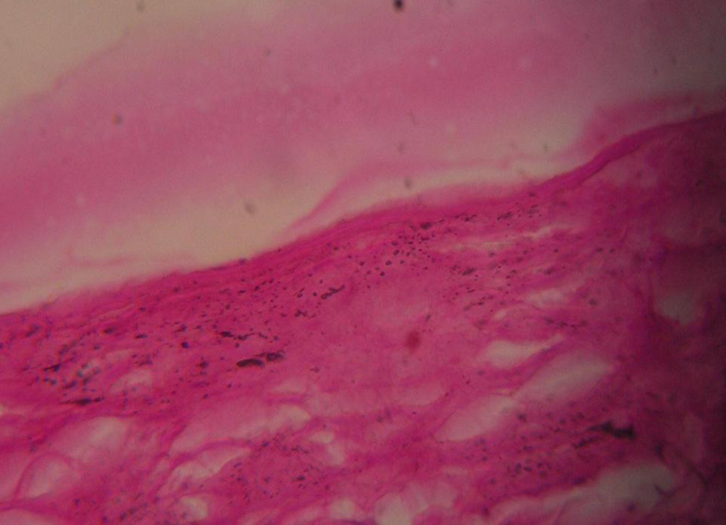
Splenic mucinous cystic tumors are defined as cystic spaces lined by mucin-producing columnar cells and ranged from benign cystadenoma to frankly malignant cystadenocarcinoma. Primary mucinous cystadenoma of spleen is an extremely rare finding [3,4,6,7]. The exact histogenetic mechanism of splenic mucinous cystic tumors is unknown except those arising from heterotopic pancreatic tissues [1]. However, mucinous cysts are usually found in association with the cystadenoma of pancreas, pseudomyxoma peritonei and mucocele of appendix [7].Only few cases, in which the tissue of origin was un-defined, are documented in the literature [3-7].
Simple mucinous cyst of spleen is a rare and close differential diagnosis, which may rarely present as pseudomyxoma peritonei [9]. Distinction is difficult to make because of low incidence and nonspecific imaging finding. Ultrasound and MRI scan, in case of mucinous cystadenoma may demonstrate a cystic lesion which may or may not contain an internal septum. Anyway, definitive diagnosis is made by surgical exploration and histopathology. Only few cases of primary mucinous cystadenoma of spleen have been reported in which tissue of origin was undefined although pseudomyxoma peritonei may uncommonly present as splenic metastasis [10]. In our case, pancreas was normal and there were no definite heterotopic pancreatic tissues in and around the splenic lesion. So, the exact histogenetic mechanism was not known. Going by Morinagas presentation [3], we concluded that the primary splenic mucinous cystadenoma may originate in the invagination of the splenic capsular mesothelium.
In conclusion,a rare case of primary mucinous cystadenoma of spleen is presented. Tissue of origin may or may not be identifiable, but it is hetertopic pancreas usually or invaginated splenic capsular mesothelium.
Acknowledgments
The author declares no conflict of interest.
| References | ▴Top |
- Zanetti G, Riccioni L, Gallo C, Salfi N, Martinelli GN. Splenic mucinous cystadenocarcinoma arising in heterotopic pancreatic tissue. Tumori. 1998;84(5):606-610.
pubmed - Souilamas MR, Khayat M, el Arbi, Soulier Y. [A rare case of proctorrhagia. Apropos of a case and review of the literature]. Ann Chir. 1993;47(3):267-269.
pubmed - Morinaga S, Ohyama R, Koizumi J. Low-grade mucinous cystadenocarcinoma in the spleen. Am J Surg Pathol. 1992;16(9):903-908.
pubmed - Nisar PJ, Zaitoun AM, Lobo DN, Rowlands BJ. Heterotopic pancreas in the spleen: malignant degeneration to mucinous cystadenocarcinoma. Eur J Gastroenterol Hepatol. 2002;14(7):793-796.
pubmed - Dedic N, Premuzic M, Cavka S, Ostojic R, Hrstic I, Vucelic B. [Pseudomyxoma peritonei associated with splenic mucinous epithelial cysts--case report]. Lijec Vjesn. 2000;122:11-12.272-275.
pubmed - Kapoor S, Naik S, Sharma S, Varshney S. Pseudomyxoma peritonei due to a ruptured mucinous cystadenoma of the spleen. European Surgery. 2007;39(5):314-316.
pubmed - Hirota M, Hayashi N, Tomioka T, Murakami S, Ohshima H, Yamasaki K, Miyamoto J,
et al . Mucinous cystadenocarcinoma of the spleen presenting a point mutation of the Kirsten-ras oncogene at codon 12. Dig Dis Sci. 1999;44(4):768-774.
pubmed - Morgenstern L, Rosenberg J, Geller SA. Tumors of the spleen. World J Surg. 1985;9(3):468-476.
pubmed - Kruslin B. [Pseudomyxoma peritonei associated with mucinous epithelial cysts of the spleen]. Lijec Vjesn. 2001;123:5-6.
pubmed - Shimoyama S, Kuramoto S, Kawahara M, Yamasaki K, Endo H, Murakami T, Kaminishi M. A rare case of pseudomyxoma peritonei presenting an unusual inguinal hernia and splenic metastasis. J Gastroenterol Hepatol. 2001;16(7):825-829.
pubmed
This is an open-access article distributed under the terms of the Creative Commons Attribution License, which permits unrestricted use, distribution, and reproduction in any medium, provided the original work is properly cited.
Journal of Clinical Medicine Research is published by Elmer Press Inc.