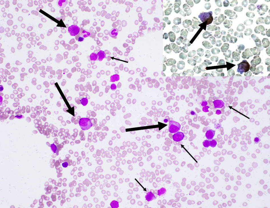
Figure 1. Bone marrow aspiration showed a hypocellular marrow with increased blasts (narrow arrows), and promyelocytes (wide arrows) with azurophil granules that were positive on peroxidase staining (upper right box) (magnification, × 400).
| Journal of Clinical Medicine Research, ISSN 1918-3003 print, 1918-3011 online, Open Access |
| Article copyright, the authors; Journal compilation copyright, J Clin Med Res and Elmer Press Inc |
| Journal website https://www.jocmr.org |
Case Report
Volume 14, Number 3, March 2022, pages 136-141
Acute Promyelocytic Leukemia in a Patient With Chronic Continuous Type of Crohn’s Disease
Figures
Tables
| Test | Value | Test | Value | Test | Value |
|---|---|---|---|---|---|
| WBC: white blood cells; RBC: red blood cells; Hb: hemoglobin; MCV: mean corpuscular volume; PLT: platelets; TP: total protein; Alb: albumin; T-Bil: total bilirubin; D-Bil: direct bilirubin; AST: aspartate aminotransferase; ALT: alanine aminotransferase; Cre: creatinine; BUN: blood urea nitrogen; LDH: lactate dehydrogenase; CRP: C-reactive protein; UIBC: unsaturated iron binding capacity; IgG: immunoglobulin G; IgA: immunoglobulin A; IgM: immunoglobulin M; APTT: activated partial thromboplastin time; PT-INR: prothrombin time international normalized ratio; Fib: fibrinogen; FDP: fibrinogen and fibrin degradation products; AT-3: antithrombin 3. | |||||
| WBC | 0.7 × 10⁹/L | TP | 7.6 g/dL | Ferritin | 10 ng/mL |
| Blasts | 1.0% | Alb | 4.6 g/dL | Vitamin B12 | 336 pg/mL |
| Promyelocytes | 0% | T-Bil | 1.6 mg/dL | Folic acid | 5.0 ng/mL |
| Myelocytes | 0% | D-Bil | 0.3 IU/L | Haptoglobin | 43 mg/dL |
| Metamyelocytes | 0% | AST | 21 IU/L | IgG | 1212 mg/dL |
| Neutrophils | 63.0% | ALT | 24 IU/L | IgA | 322 mg/dL |
| Eosinophils | 0% | Cre | 0.77 mg/dL | IgM | 62 mg/dL |
| Basophils | 0% | BUN | 12.3 mg/dL | APTT | 28.1 s |
| Monocytes | 0% | LDH | 140 IU/L | PT-INR | 0.99 |
| Lymphocytes | 36% | Na | 145 mEq/L | Fib | 224 mg/dL |
| RBC | 3.15 × 10¹²/L | K | 3.8 mEq/L | FDP | 37.7 mg/dL |
| Hb | 10.9 g/dL | Cl | 108 mEq/L | D-dimer | 14.5 µg/mL |
| MCV | 101.9 fL | CRP | 0.27 mg/dL | AT-3 | 96.6% |
| Reticulocytes | 19‰ | Fe | 70 µg/dL | ||
| PLT | 154 × 109/L | UIBC | 469 µg/dL | ||
| N | Agea (years) | Durationb | Treatment for Crohn’s disease | WBC (/µL) | Hb (g/dL) | PLT (× 104/µL) | Inflammatory biomarkerc | G-banding analysis | Outcome | Reference |
|---|---|---|---|---|---|---|---|---|---|---|
| aAge at diagnosis of APL. bDuration from diagnosis of Crohn’s disease to APL. cValues in the parenthesis mean the highest and worst values of the inflammatory markers at the onset of APL. WBC: white blood cell; Hb: hemoglobin; PLT: platelet; 5-ASA: 5-aminosalicylic acid; IFX: infliximab; PML-RARA: promyelocytic leukemia/retinoic acid receptor α; FISH: fluorescence in situ hybridization; CR: complete response; NA: not available; ESR: erythrocyte sedimentation rate; CRP: C-reactive protein. | ||||||||||
| 1 | 42 | Long time | Steroids, sulfasalazine, 6-mercaptopurine, azathioprine, IFX | 1,700 | 7.6 | Normal | NA | Normal karyotype (PML-RARA FISH positive) | CR > 5 years | [11] |
| 2 | 13 | 9 months | Steroids, 5-ASA | NA | 6.5 | NA | NA | NA | CR 1 year | [12] |
| 3 | 78 | 11 years | Steroids, 5-ASA | 4,600 | 6.8 | 6.9 | ESR (120 mm/h), ferritin (1,200 ng/mL) | NA | Died < 1 month | [13] |
| 4 | 37 | 8 years | Steroids, 5-ASA | 28,500 | NA | NA | ESR, CRP (normal values) | NA | Alive 2 years | [13] |
| 5 | 49 | 23 years | Multiple small bowel resections, colostomy | 600 | 11 | 3.3 | NA | NA | NA | [14] |
| 6 | 19 | 2 years | Sulfasalazine | 1,400 | NA | 6.4 | NA | t(15;17)/+8,t(15;17) | Died < 1 year | [15] |
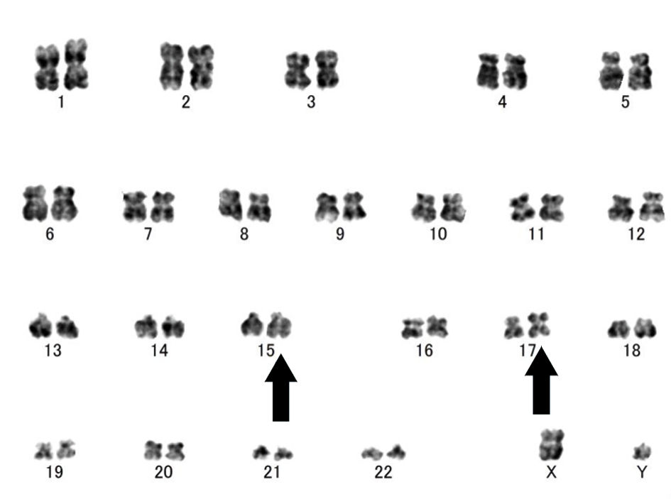
| Case in this article | 51 | 23 years | 5-ASA, steroids, IFX, ustekinumab | 700 | 10.9 | 15.4 | CRP (0.27 mg/dL) | ider(17)(q10)t(15;17)/der(15)t(15;17)(q24.1: q21.2) | CR | |