Figures
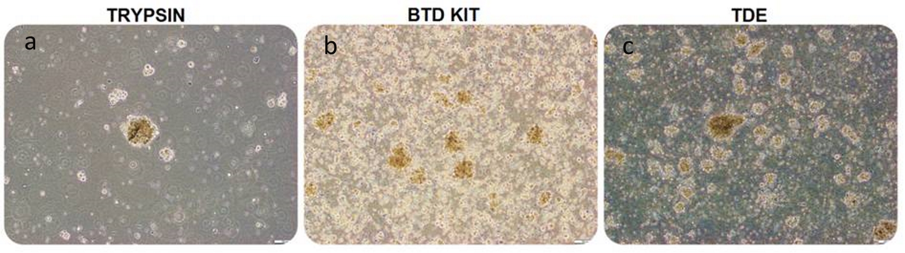
Figure 1. Glial tumor stem cells, isolated with trypsin, BTD Kit and TDE from equal amount of glioblastoma specimen, seeded 1 × 10⁶ cells into each flask were visualized by phase-contrast microscopy on day 4, passage 0 in suspension culture (bars: a-b:100 µm, c: 200 µm). Microscopic observation revealed that the number of spheres formed by trypsin-isolation (a) is lower than the other groups. The number of spheres formed by BTD Kit-isolation (b) is close to the number of spheres formed by TDE-isolated (c). BTD Kit: brain tumor dissociation Kit; TDE: tumor dissociation enzyme.
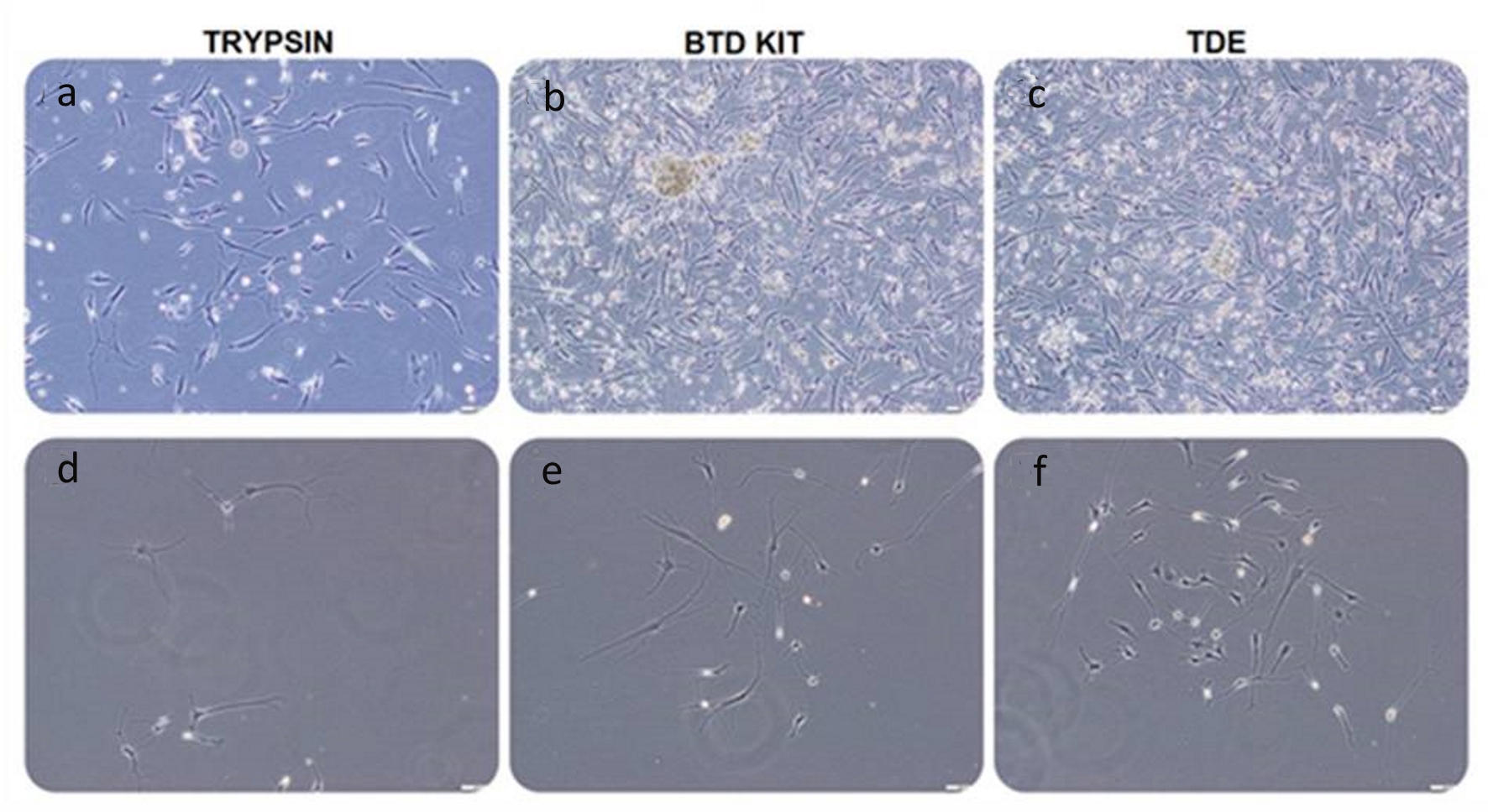
Figure 2. Glial tumor stem cells, isolated with trypsin, BTD Kit and TDE from equal amount of GBM specimen, seeded 1 × 10⁶ cells into each flask were visualized by phase-contrast microscopy on day 12 (a-c) and day 4 (d-f), passage 0 in adherent culture (bars: 100 µm). Microscopic observation revealed that the number of adherent glial tumor stem cells isolated with trypsin (a, d) is less than that of other groups. The number of cells isolated with BTD Kit (b, e) is close to the number of cells isolated with TDE (c, f). BTD Kit: brain tumor dissociation Kit; TDE: tumor dissociation enzyme.
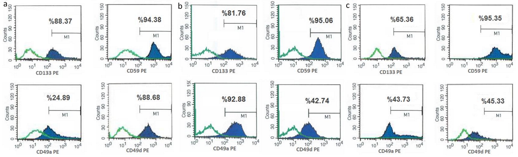
Figure 3. Figure 3. Flow cytometry analyzes of glial tumor stem cells (isolated with trypsin (a), BTD Kit (b) and TDE (c)) with glial tumor stem cell markers (CD133, CD59, CD49a, CD49d). Flow cytometry analysis of collected cells from culture dishes was performed. Positive expressions of CD133, CD59, CD49a and CD49d indicate that isolated and analyzed tumor stem cells have GBM characteristics. These characteristics of isolated tumor stem cells by three different methods were compared. CD133 and CD59e expressions were found to be similar in all cells, whereas expression of CD49a was higher in cells isolated with BTD Kit, and expression of CD49d was higher in cells isolated with trypsin. This shows that these isolation methods with BTD Kit and trypsin are more successful to isolate cells which have higher expression of glial tumor stem cell markers. BTD Kit: brain tumor dissociation Kit; TDE: tumor dissociation enzyme.
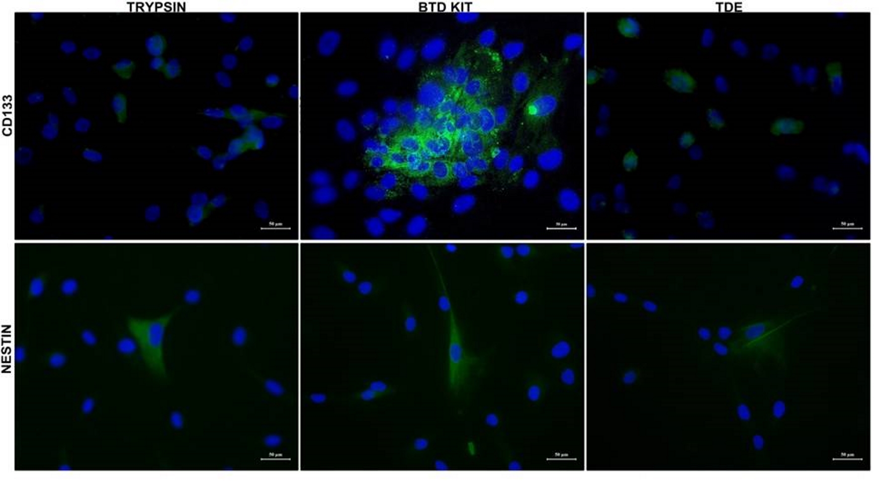
Figure 4. CD133 and nestin expression in glial tumor stem cells. Glial tumor stem cells were cultured with growth medium in chambered cell culture slides. After adhering in culture slides, cells were fixed and stained to detect CD133 and nestin. Immunofluorescence microscopy imaging of the expression of CD133 and nestin in isolated cells by three different methods are shown. Nuclei were stained with DAPI (blue) (bars: 50 µm). DAPI: 4’,6-diamidino-2-phenylindole.
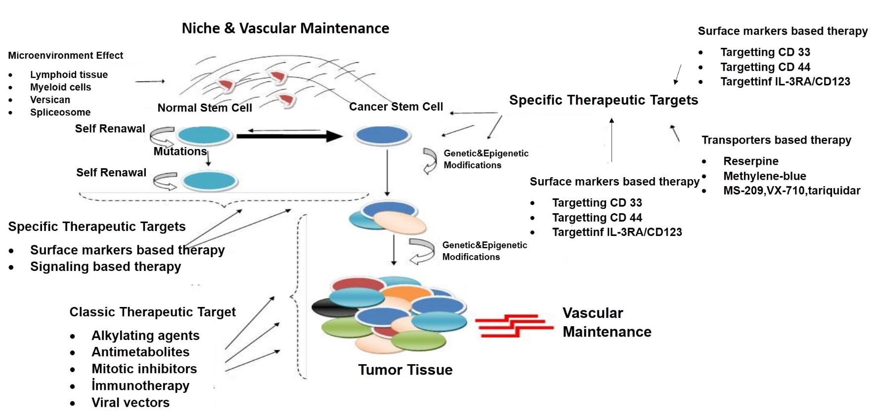
Figure 5. New therapeutic impact areas in cancer stem cell theory. This figure has showed many recent approaches to stem cell such as targeting of cell surface molecules, cell penetrating peptides, immunotherapy and change of microenvironment of niche.