Journal of Clinical Medicine Research, ISSN 1918-3003 print, 1918-3011 online, Open Access
Article copyright, the authors; Journal compilation copyright, J Clin Med Res and Elmer Press Inc
Journal website http://www.jocmr.org

Case Report

Volume 11, Number 1, January 2019, pages 72-75


Persistent Primitive Hypoglossal Artery Associated With Multiple Cerebral Aneurysms

Figures

Figure 1.
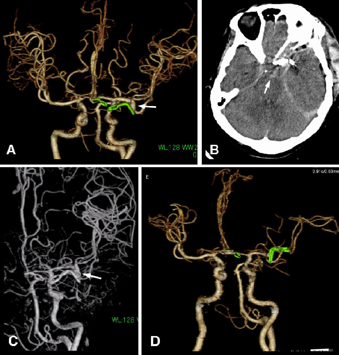
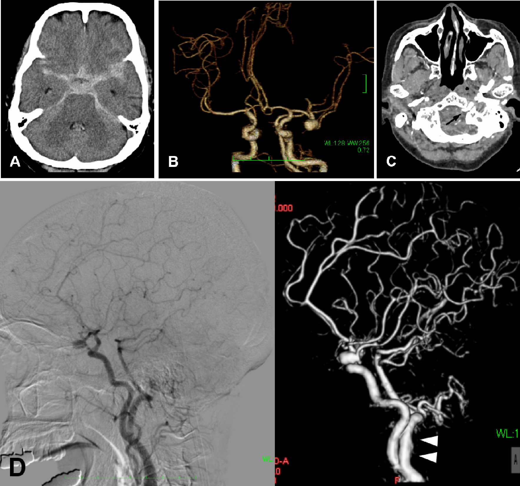
Figure 1. (A) CT demonstrating SAH and enlargement of the ventricles. (B) 3D-CTA demonstrating two aneurysms at AComA and the left MCA. The left MCA aneurysm projects medially. (C) A raw image of CTA showing an artery running through the left hypoglossal canal (arrow). (D) Angiography (left: conventional, right: 3D) demonstrating PPHA (arrowheads) originating from the left ICA.
Figure 2.
Figure 2. (A) Post-operative 3D-CTA demonstrating a small residual neck of the MCA aneurysm (arrow). (B) CT on the sixth day showing additional hemorrhage in the basal cistern (arrow). (C) Angiography showing enlargement of the residual neck of the MCA aneurysm (arrow). (D) After the second operation, the aneurysm is not observed on 3D-CTA.