Journal of Clinical Medicine Research, ISSN 1918-3003 print, 1918-3011 online, Open Access
Article copyright, the authors; Journal compilation copyright, J Clin Med Res and Elmer Press Inc
Journal website http://www.jocmr.org

Case Report

Volume 9, Number 11, November 2017, pages 962-964


Newly Diagnosed Idiopathic Liver Abscess: Colonoscopy Required!

Figures

Figure 1.
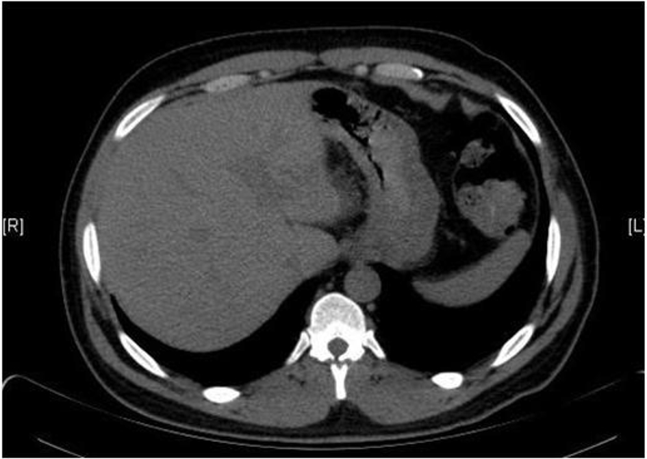
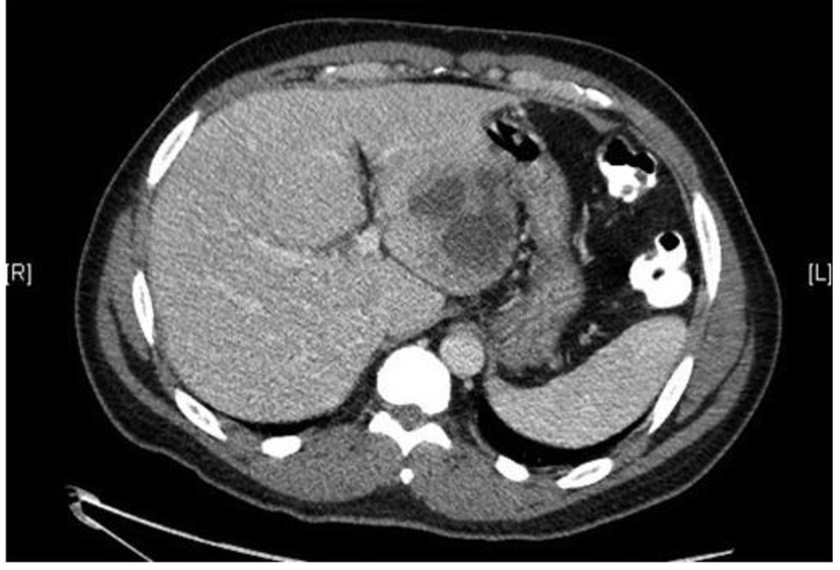
Figure 1. CT of abdomen/pelvis with contrast showing 7.5 × 7.0 cm multilobulated complex mass in the left lobe of the liver.
Figure 2.
Figure 2. Diagnostic colonoscopy revealing a 5 cm pedunculated polyp in the sigmoid colon.
Figure 3.
Figure 3. Pathology of the sigmoid colon polyp showing moderately differentiated invasive adenocarcinoma.
Figure 4.
Figure 4. Liver biopsy showing granulation tissue surrounded by fibrotic cap; consistent with an abscess.
Figure 5.
Figure 5. CT of abdomen/pelvis with contrast after 3 weeks of antibiotic therapy showing an interval decrease in size of the left lobe hepatic mass, now measuring 3.6 × 3.0 cm.