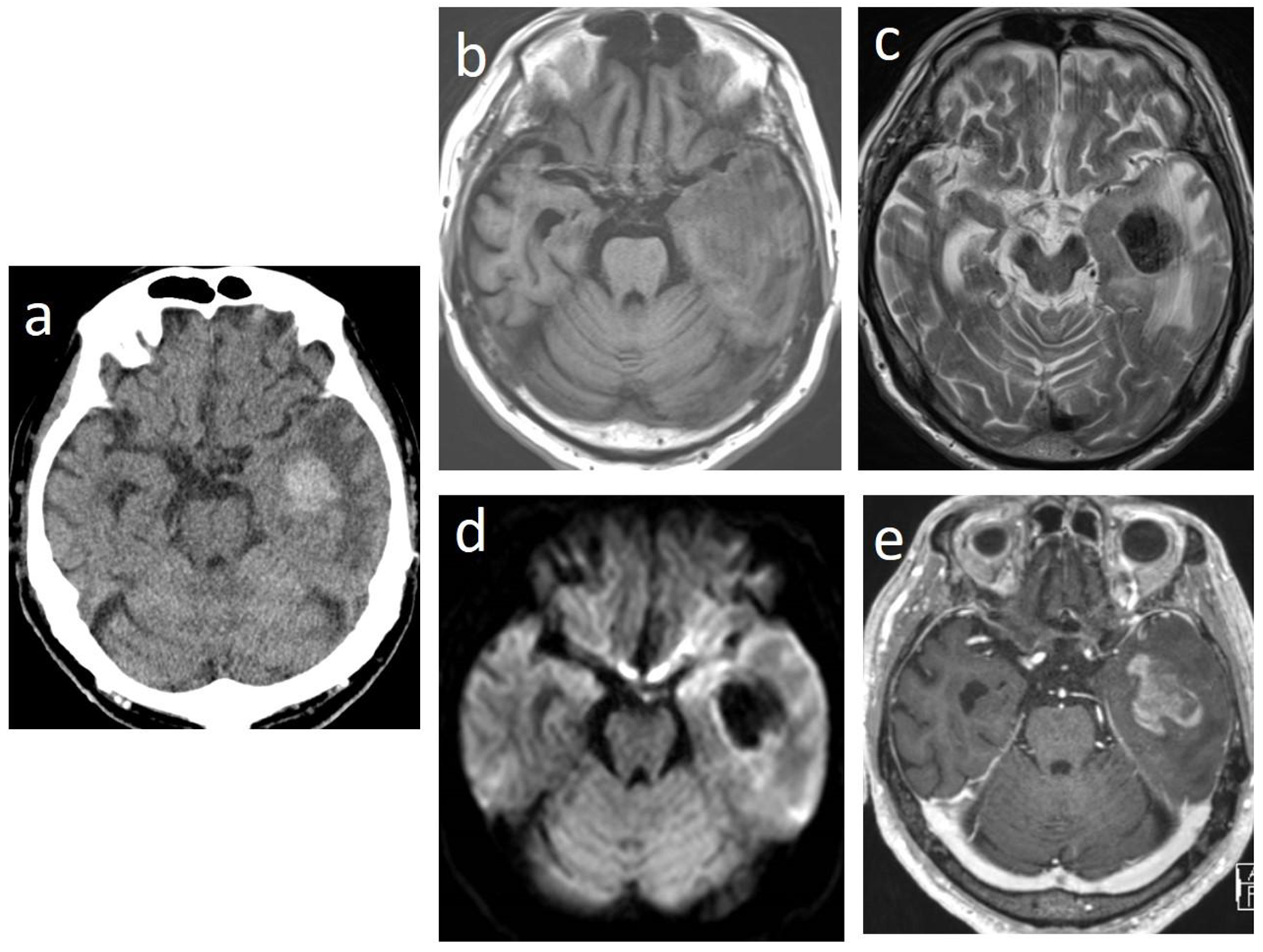
Figure 1. Brain computed tomographic images showing a high-density tumor with edema and intratumoral hemorrhage in the left temporal lobes (a). The tumor is iso- to hypointense on T1-weighted magnetic resonance imaging (MRI) (b) and hypointense on T2-weighted MRI (c) and on diffusion-weighted MRI (d). Gadolinium-enhanced T1-weighted MRI showing heterogeneous enhancement (e).
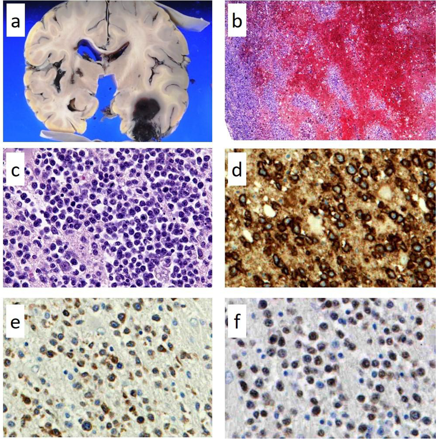
Figure 2. Pathological findings of autopsy in the left temporal lobe. Focal hemorrhages in the left temporal lobe (a) in which lymphoma cells had diffusely infiltrated with hemorrhage; (b) hematoxylin and eosin staining × 100; (c) × 400. The lymphoma cells in the brain lesion are positive for CD20 (d, × 400); CD79a (e, × 400); and PAX5 (f, × 400).
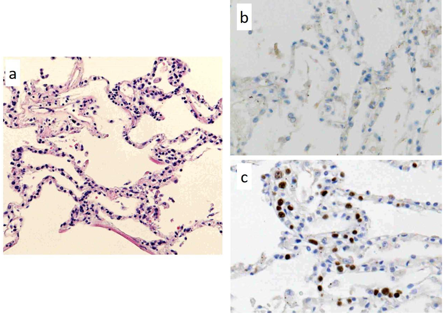
Figure 3. Pathological findings of autopsy in a part of the lung lesions. There are lymphoma cells within the lumina of vessels; (a) hematoxylin and eosin staining (× 100). The lymphoma cells in the lung lesion are negative for CD20 (b, × 400), but positive for PAX5 (c, × 400).