Journal of Clinical Medicine Research, ISSN 1918-3003 print, 1918-3011 online, Open Access
Article copyright, the authors; Journal compilation copyright, J Clin Med Res and Elmer Press Inc
Journal website http://www.jocmr.org

Case Report

Volume 11, Number 4, April 2019, pages 305-308


Complete Occlusion of Bilateral Internal Carotid Artery in a Marijuana Smoker: A Case Report

Figures

Figure 1.
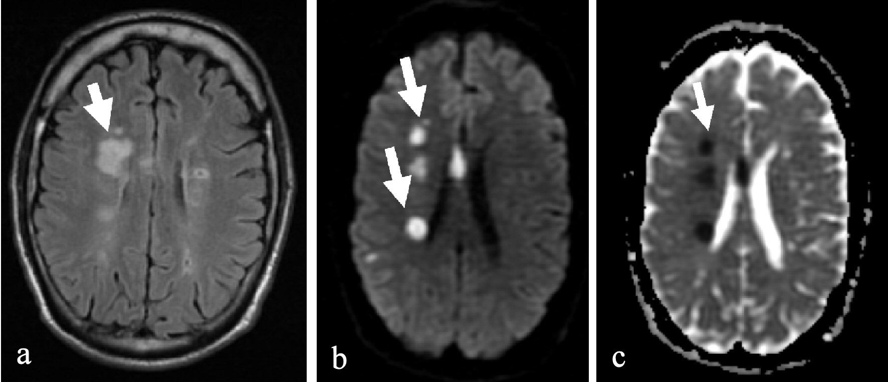
Figure 1. MRI head showed multiple foci of abnormal signal intensity in the right cerebral hemisphere and a small focus in the left parietal region representing acute ischemic changes as shown by arrow. (a) T2 flair. (b) Diffusion weighted image. (c) Apparent diffusion coefficient.
Figure 2.
Figure 2. CT angiography of the head and neck with 125 mL Optiray 350 contrast enhanced 3D volume rendered image, with white arrowheads showing ICA occlusions.
Figure 3.
Figure 3. CT angiography of the head with 125 mL Optiray 350 contrast, with white arrowheads showing the ICA occlusions with calcification. IJ: internal jugular.