Journal of Clinical Medicine Research, ISSN 1918-3003 print, 1918-3011 online, Open Access
Article copyright, the authors; Journal compilation copyright, J Clin Med Res and Elmer Press Inc
Journal website http://www.jocmr.org

Case Report

Volume 9, Number 10, October 2017, pages 886-888


A Rare Case of Atrial Metastasis From a Rectal Adenocarcinoma

Figures

Figure 1.
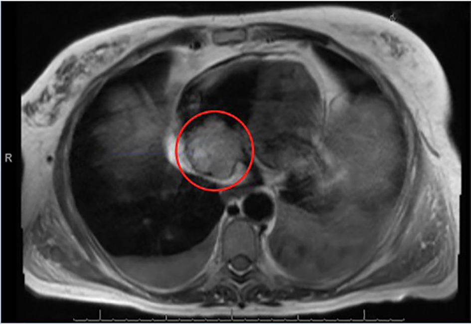
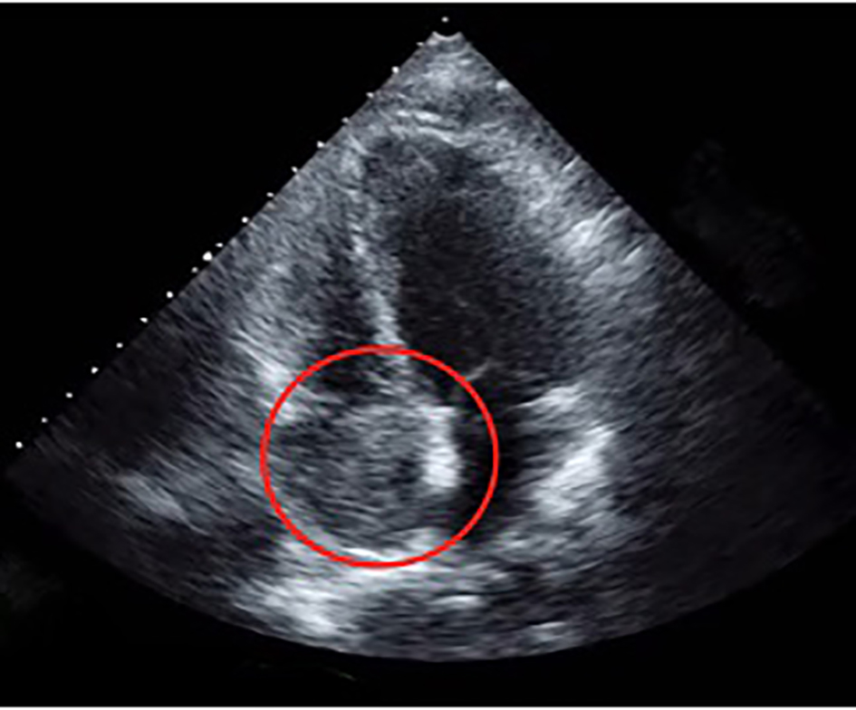
Figure 1. A transesophageal echocardiogram in a four-chamber view showing a right atrial mass, 4 × 3.5 cm.
Figure 2.
Figure 2. A T2-weighted MRI showing the right atrial, non-mobile and heterogeneously hyperintense mass, measuring 4.6 × 4.2 cm.
Figure 3.
Figure 3. Sagittal oblique T2-weighted MRI of the chest showing a heterogeneously hyperintense filling defect in the inferior vena cava from the level of the renal veins into the right atrium. No hepatic mass was present.
Figure 4.
Figure 4. Sagittal oblique T2-weighted sequence through the right heart inflow and outflow tracts showing the right atrial mass in the right atrium.