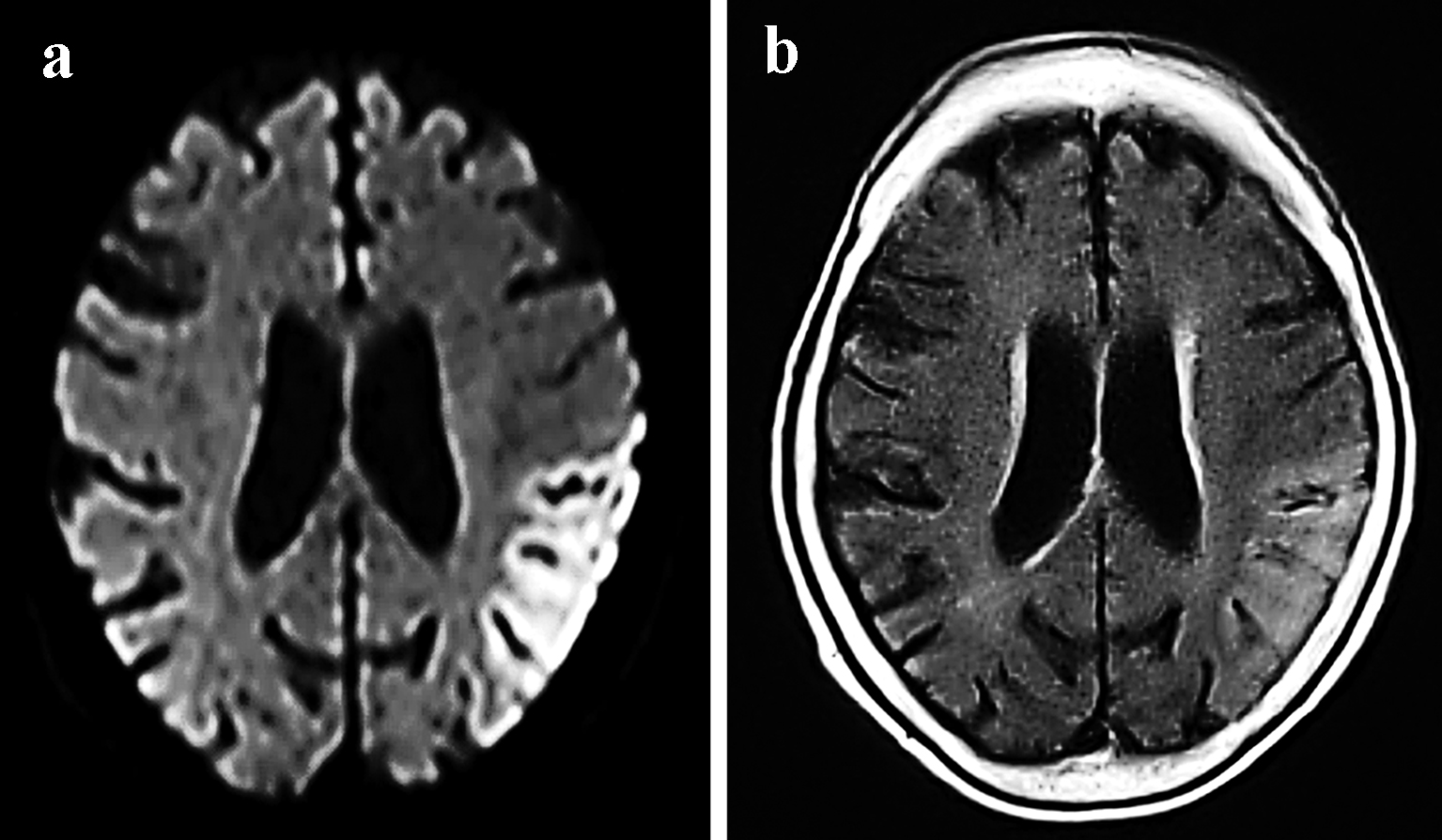
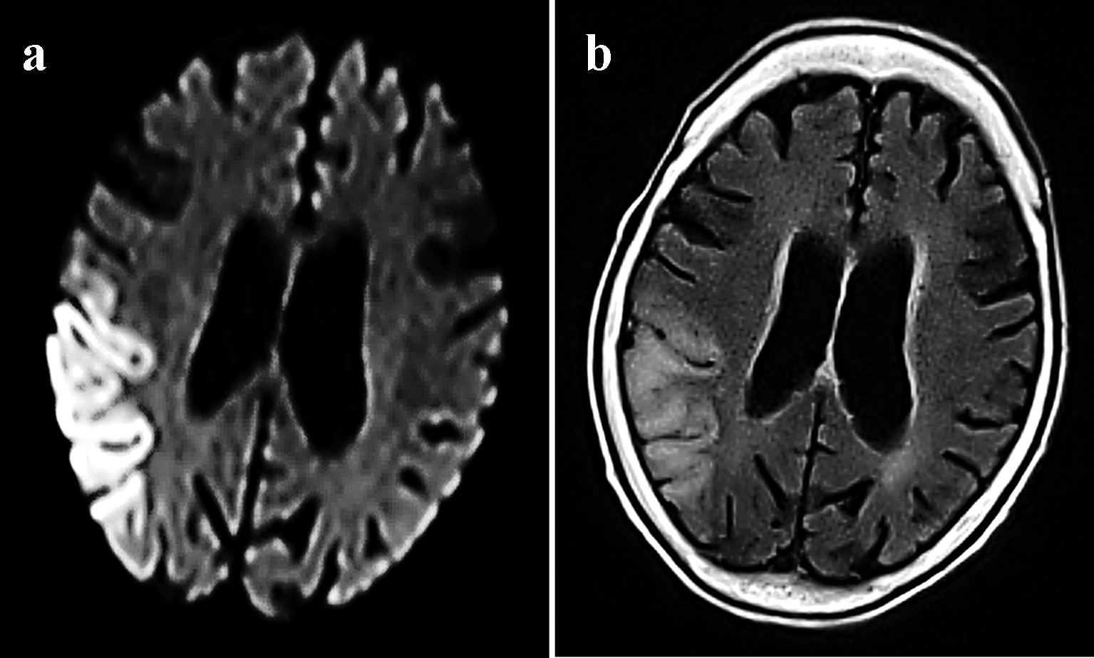
Figure 1. MRI examinations from May 2014 showing the migratory lesions of mitochondrial encephalomyopathy, lactic acidosis, and stroke-like episodes. Cortical DWI (a) and FLAIR (b) sequences revealed hyperintensity in the left temporal lobe.
| Journal of Clinical Medicine Research, ISSN 1918-3003 print, 1918-3011 online, Open Access |
| Article copyright, the authors; Journal compilation copyright, J Clin Med Res and Elmer Press Inc |
| Journal website http://www.jocmr.org |
Case Report
Volume 9, Number 9, September 2017, pages 812-819
Importance of Distinguishing Between Mitochondrial Encephalomyopathy With Elderly Onset of Stroke-Like Episodes and Cerebral Infarction
Figures
Tables
| Age at diagnosis (years) | Sex | Main clinical features | Neuroimaging findings | Mutation | Reference |
|---|---|---|---|---|---|
| DM: diabetes mellitus. | |||||
| 67 | F | Seizures, stroke-like episodes, hearing deficit, progressive higher brain function impairment and DM | Bilateral temporal lobe and basal ganglia calcification | m.3243A>G | Our case |
| 70 | F | Headaches, hearing deficit, DM and encephalopathy | Right temporal and basal ganglia calcification | m.3243A>G | Aurangzeb et al, 2014 [5] |
| 66 | F | Encephalopathy, proximal myopathy and DM | Left periventricular lacunar infarction and prominent calcification of the pineal gland and basal ganglia | m.3243A>G | Jones et al, 2004 [6] |
| CT: computed tomography; MRI: magnetic resonance imaging. |
| Recurrent stroke-like episodes |
| Head scans (CT and MRI) that are inconsistent with the main blood-vessel-dominant region |
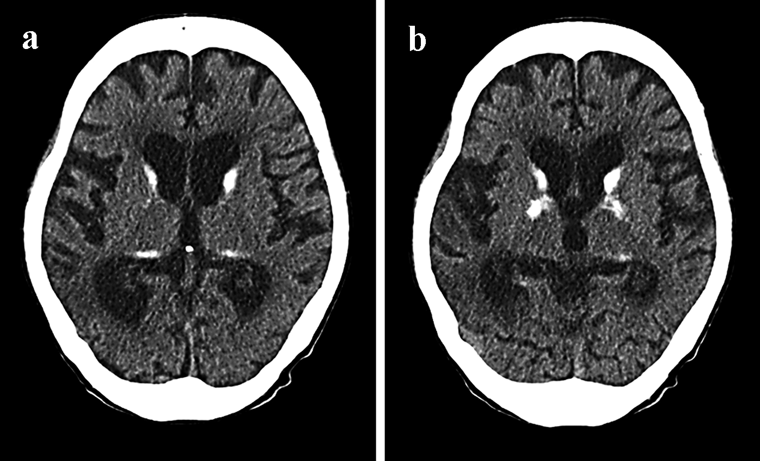
| CT scans revealing calcification around the basal ganglia |
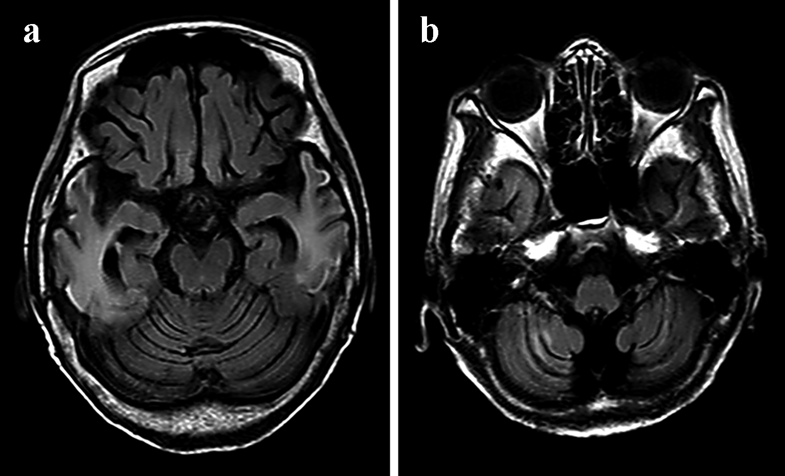
| Atrophy of the cerebrum |
| Sensorineural hearing loss from a young age |
| Progressive higher brain dysfunction |
| Emaciation |
| Presence of a family history |
| Recurrent seizures |
| Disturbances of consciousness |