Journal of Clinical Medicine Research, ISSN 1918-3003 print, 1918-3011 online, Open Access
Article copyright, the authors; Journal compilation copyright, J Clin Med Res and Elmer Press Inc
Journal website http://www.jocmr.org

Review

Volume 8, Number 8, August 2016, pages 569-574


Estimating Right Atrial Pressure Using Ultrasounds: An Old Issue Revisited With New Methods

Figures

Figure 1.
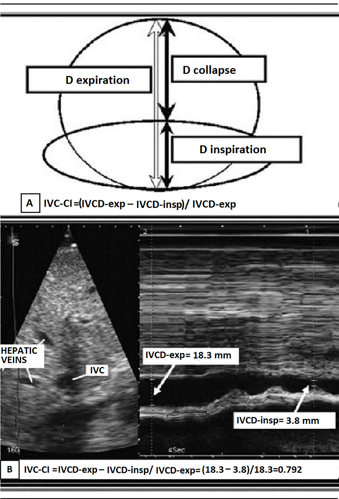
Figure 1. (A) Representation of the IVC collapsibility index (IVCCI) and (B) IVCCI measurement using M-mode ultrasonography. (A) IVCCI consists of the difference between the end-expiratory (IVCd-exp) and end-inspiratory (IVCd-insp) divided by IVCd-exp. (B) Based on the measurements in this example, the IVCCI would be (18.3 - 3.8 mm)/18.3 mm, or 79.2%.
Figure 2.
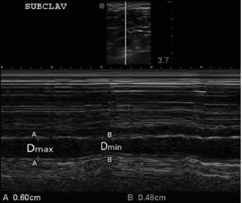
Figure 2. B-mode and M-mode views of the subclavian vein (SCV) with expiratory and inspiratory diameters measured. The clavicle is visualized anterior (above) the subclavian artery at the left side of the top image (superior to the SVC). Again, consistent with our approach to the IVCCI, we utilized minimal (inspiratory) and maximal (expiratory) diameters of the SCV. For further explanations, please see the text.

Tables

Table 1. Hemodynamic Monitoring With a Pulmonary Artery Catheter: Normal Pressures and Resistance Values
 
MeanRange
Right atrium4 mm Hg1 - 8 mm Hg
Right ventricle
Peak-systolic25 mm Hg15 - 30 mm Hg
End-diastolic9 mm Hg4 - 12 mm Hg
Pulmonary capillary wedge pressure9 mm Hg4 - 12 mm Hg
Systemic vascular resistance1,100 dyne-s/cm5700 - 1,600 dyne-s/cm5
Pulmonary vascular resistance70 dyne-s/cm520 - 130 dyne-s/cm5


Table 2. Estimation of RA Pressure on the Basis of IVC Diameter and Collapse According to Rudski et al [19]
 
VariablesNormal (0 - 5(3) mm Hg)Intermediate (5 - 10(8) mm Hg)High (15 mm Hg)
Ranges are provided for low and intermediate categories, but for simplicity, midrange values of 3 mm Hg for normal and 8 mm Hg for intermediate are suggested. Intermediate (8 mm Hg) RAPs may be downgraded to normal (3 mm Hg) if no secondary indices of elevated RAP are present, upgraded to high if minimal collapse with sniff (< 35%) and secondary indices of elevated RAP are present, or left at 8 mm Hg if uncertain. IVC: inferior vena cava; RAP: right atrial pressure. The table synthetically displays the concepts expressed by Rudski et al [19] in the official recommendations of the American Society of Echocardiography (2010). These criteria have been left unchanged in the recent update [20] (January 2015).
IVC diameter≤ 21 mm≤ 21 mm; > 21 mm> 21 mm
Collapse with sniff> 50%< 50%; > 50%< 50%
Secondary indices of elevated RAPRestrictive filling
Tricuspid E/e’ > 6
Diastolic flow predominance in hepatic veins (systolic filling fraction < 55%)


Table 3. Accuracy, Sensitivity and Specificity of Echocardiographic Measurements for Identification of RAP > 10 mm Hg According to Patel et al [21]
 
3D-RAVi ≥ 35 mL/m2 + IVC ≥ 2 cm3D-RAVi ≥ 35 mL/m23D-RAVi ≥ 35 mL/m2 + IVC ≥ 2 cm + IVCCI < 40%IVC ≥ 2 cmIVC ≥ 2 cm + IVCCI < 40%
Comparison of 3D-RAVi ≥ 35 mL/m2 + IVC ≥ 2 cm versus IVC parameters alone. *P = 0.038 versus IVC ≥ 2 cm + IVCCI < 40%; †P = 0.041 vs. IVC ≥ 2 cm + IVCCI < 40%. RAP: right atrial pressure; 3D-RAVi: three-dimensional right atrial volume index; IVC: inferior vena cava expiratory diameter; IVCCI: inferior vena cava collapsibility index.
Accuracy0.88*0.850.700.830.68
Sensitivity0.86†0.890.570.890.60
Specificity0.920.751.000.670.83