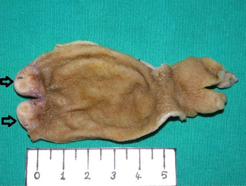
Figure 1. A white, firm hemisferic mass containing microcysts in the fundal region of the gallbladder (black arrows).
| Journal of Clinical Medicine Research, ISSN 1918-3003 print, 1918-3011 online, Open Access |
| Article copyright, the authors; Journal compilation copyright, J Clin Med Res and Elmer Press Inc |
| Journal website http://www.jocmr.org |
Case Report
Volume 2, Number 3, June 2010, pages 150-153
Fundal Variant Adenomyomatosis of the Gallbladder: Report of Three Cases and Review of the Literature
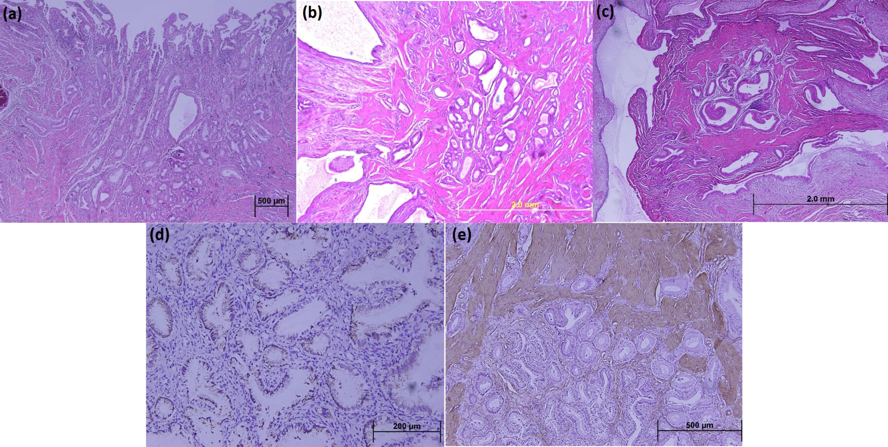
Figures