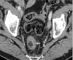
Figure 1. Transversal view of abdominal CT scan. Circumferential thickening of the rectal wall (arrow).
| Journal of Clinical Medicine Research, ISSN 1918-3003 print, 1918-3011 online, Open Access |
| Article copyright, the authors; Journal compilation copyright, J Clin Med Res and Elmer Press Inc |
| Journal website http://www.jocmr.org |
Case Report
Volume 2, Number 3, June 2010, pages 137-139
Rectal Metastasis of Prostate Cancer: About a Case
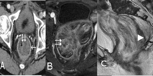
Figures