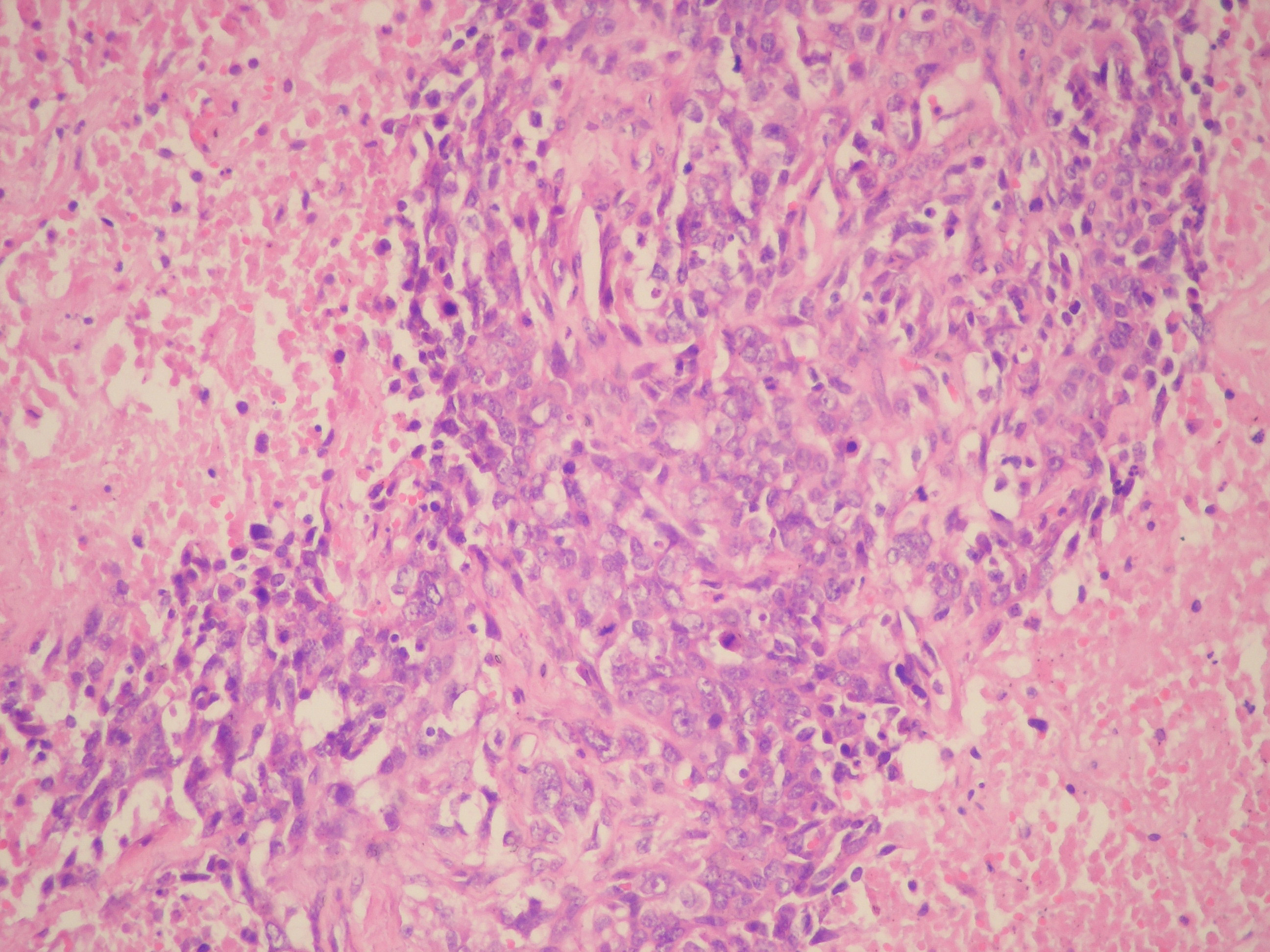
Figure 1. Circular tumor areas containing focally necrosis and infiltration of adipose tissue were seen (HE x 40).
| Journal of Clinical Medicine Research, ISSN 1918-3003 print, 1918-3011 online, Open Access |
| Article copyright, the authors; Journal compilation copyright, J Clin Med Res and Elmer Press Inc |
| Journal website http://www.jocmr.org |
Case Report
Volume 2, Number 2, April 2010, pages 96-98
A Rare Tumour of the Breast: Carcinosarcoma
Figures