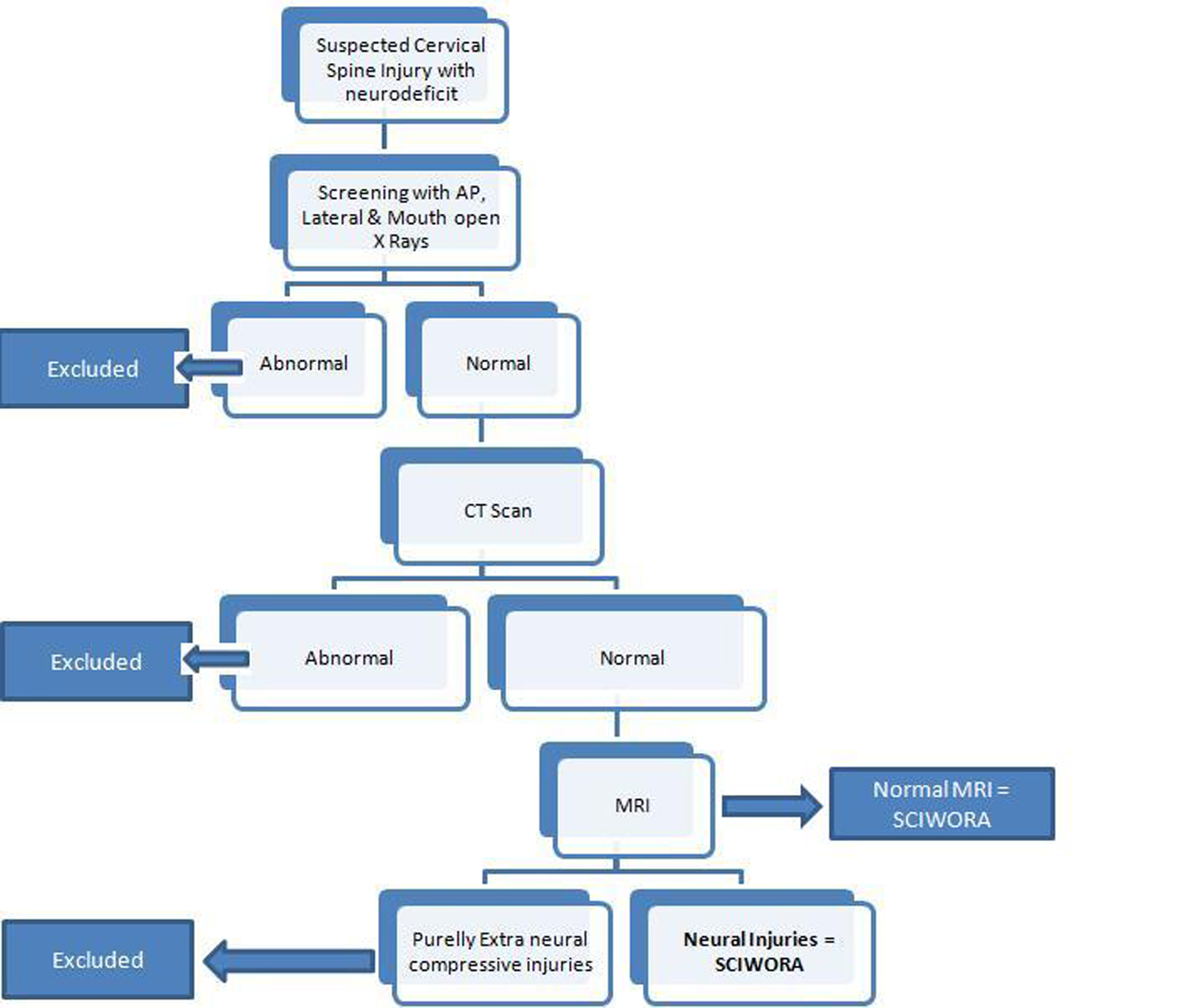
Figure 1. Algorithm for diagnosis of adult SCIWORA.
| Journal of Clinical Medicine Research, ISSN 1918-3003 print, 1918-3011 online, Open Access |
| Article copyright, the authors; Journal compilation copyright, J Clin Med Res and Elmer Press Inc |
| Journal website http://www.jocmr.org |
Original Article
Volume 1, Number 3, August 2009, pages 165-172
Adult Spinal Cord Injury without Radiographic Abnormalities (SCIWORA): Clinical and Radiological Correlations
Figures
Table
| Sno. | Age/Sex | Mode of Trauma | Neurological Status at Admission (AIS Grade) | Radiographs & CT Scan | Neural Injury (On MRI) | Extra Neural Injury (On MRI) | Treatment | Neurological Status at 12 months (AIS Grade) |
|---|---|---|---|---|---|---|---|---|
| MVA: Motor vehicle accident; WRA: Work related accident; CSLP: Cervical Spine Locking Plate. | ||||||||
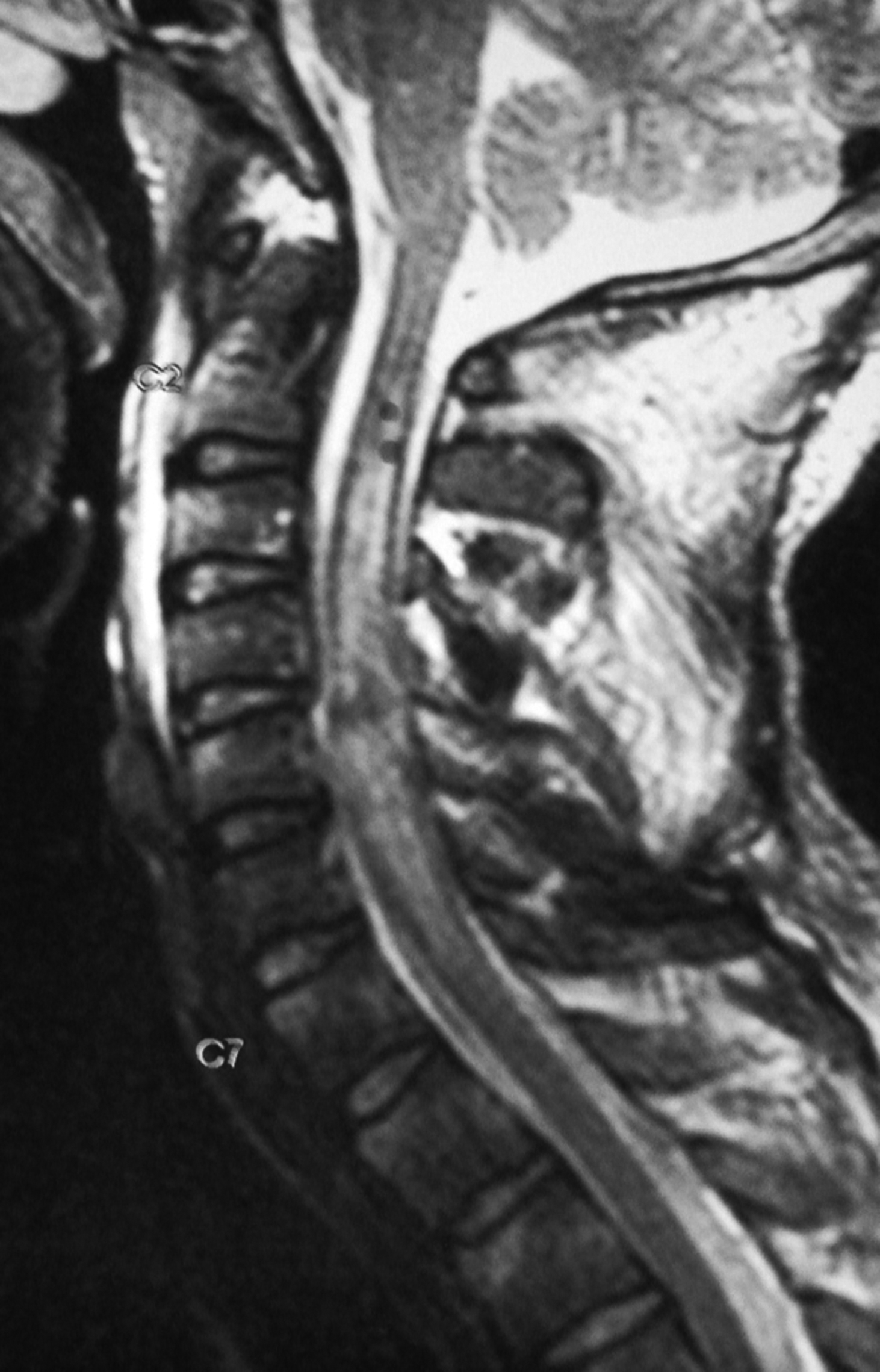
| 1. | 22 M | MVA | AIS ‘A’ | Normal | Cord contusion opposite C6,7 Pattern III | Disc bulges at C 3-4 & C6-7, L. Flavum bulging C6-7. | Conservative | AIS ‘C’ |
| 2. | 32 M | Roof Collapse | AIS ‘C’ | Normal | Cord contusion with long segment cord edema C2-7 Pattern III | L. Flavum bulge C4-5. | Conservative | AIS ‘D’ |
| 3. | 55 M | MVA | AIS ‘B’ | Spondylosis | Cord Hemorrhage. C5-6 Pattern I | Disc Prolapse C5-6 & C6-7. L.Flavum bulging from C 5-7. | Discectomy C5-6, C6-7 and Fusion. (Tricortical Iliac Graft & 2 level CSLP) | AIS ‘C’ |
| 4. | 51 M | MVA | AIS ‘C’ Central Cord Syndrome | Spondylosis | Cord edema C1-2. Pattern II | Multiple level disc bulges, L. Flavum bulge C4-5. | Conservative | AIS ‘D’ |
| 5. | 58 M | Fall from height | AIS ‘C’ | Spondylosis | Cord edema C4-6 Pattern II | - | Conservative | AIS ‘E’ |
| 6. | 33 F | Fall from Height | AIS ‘C’ | Normal | Cord edema C5-6 Pattern II | - | Conservative | AIS ‘E’ |
| 7. | 29 M | WRA | AIS ‘B’ | Spondylosis | Cord Contusion C6-7 Pattern III | Multiple Level Disc Bulges | Conservative | AIS’D’ |
| 8. | 40 M | Fall from height | AIS ‘C’ Central Cord Syndrome | Normal | Cord Contusion C4-6 Pattern III | - | Conservative | AIS ‘D’ |
| 9. | 52 F | MVA | AIS ‘C’ | Spondylosis | Cord edema C5-6 Pattern II | Disc Prolapse C5-6. | Discectomy C5-6 & Fusion (Tricortical iliac graft &1 level CSLP) | AIS ‘D’ |
| 10. | 38 M | MVA | AIS ‘A’ | Normal | Cord Hemorrhage C4-6 Pattern I | - | Conservative | AIS ‘B’ |
| 11. | 26 M | WRA | AIS ‘B’ | Normal | Cord Contusion C4-5 Pattern III | - | Conservative | AIS ‘D’ |
| 12. | 28 M | Fall from Height | AIS ‘C’ | Normal | Cord edema C4-6 Pattern II | - | Conservative | AIS ‘E’ |