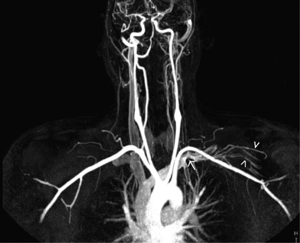
Figure 1. Coronal maximum-intensity-projection contrast-enhanced MR angiogram obtained after left antecubital vein injection reveals near-complet occlusion of the left proksimal subclavian vein (arrow) and distal venous collaterals( arrowhead).
| Journal of Clinical Medicine Research, ISSN 1918-3003 print, 1918-3011 online, Open Access |
| Article copyright, the authors; Journal compilation copyright, J Clin Med Res and Elmer Press Inc |
| Journal website http://www.jocmr.org |
Case Report
Volume 1, Number 3, August 2009, pages 178-180
Subclavian Vein Thrombosis Extending into the Internal Jugular Vein: Paget-von Schroetter Syndrome
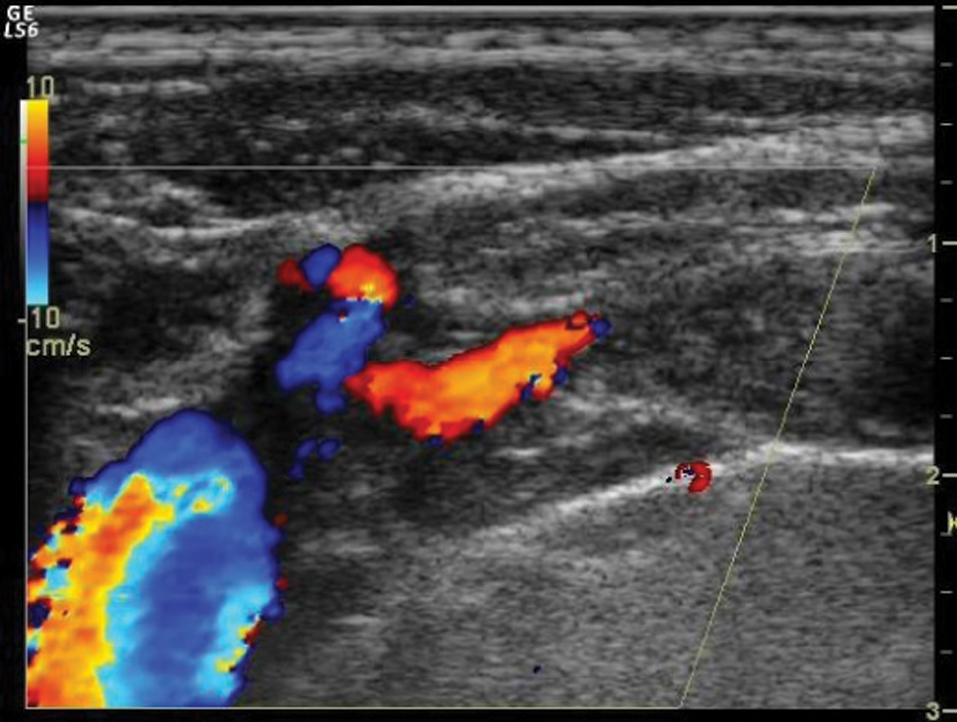
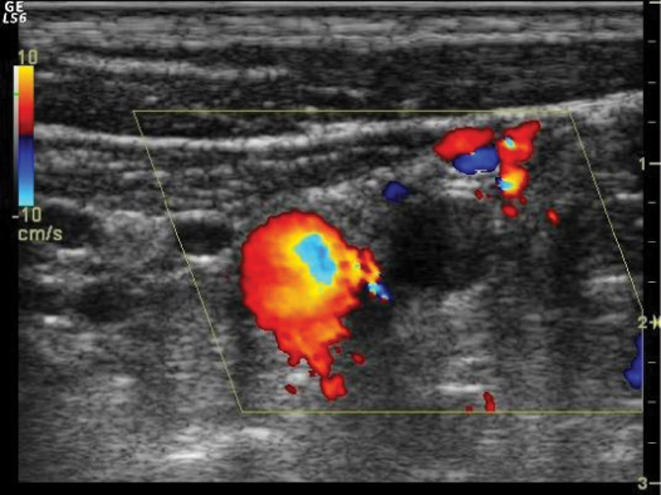
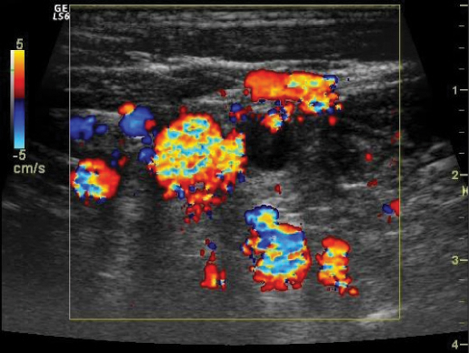
Figures