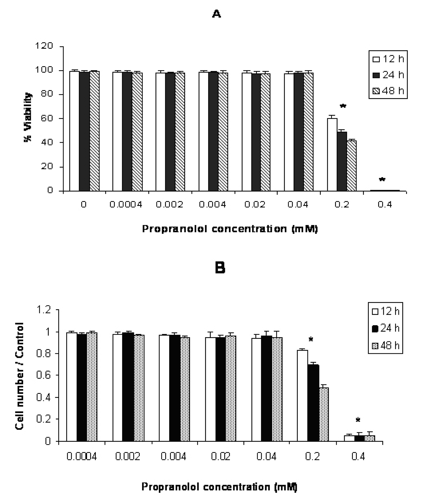
Figure 1. Effect of propranolol on proliferative responses of human leukemic Molt-4 T-cell line. The Molt-4 cells were treated with different concentrations of propranolol (0.0004 - 0.4 mM) for 12, 24 and 48 hours. The results are presented as % of viability demonstrated by trypan blue dye exclusion (TB) test (A) and cell number/control demonstrated by MTT assay (B). Data are mean ± SD of triplicate cultures. n = 3; P < 0.05 was considered significant.
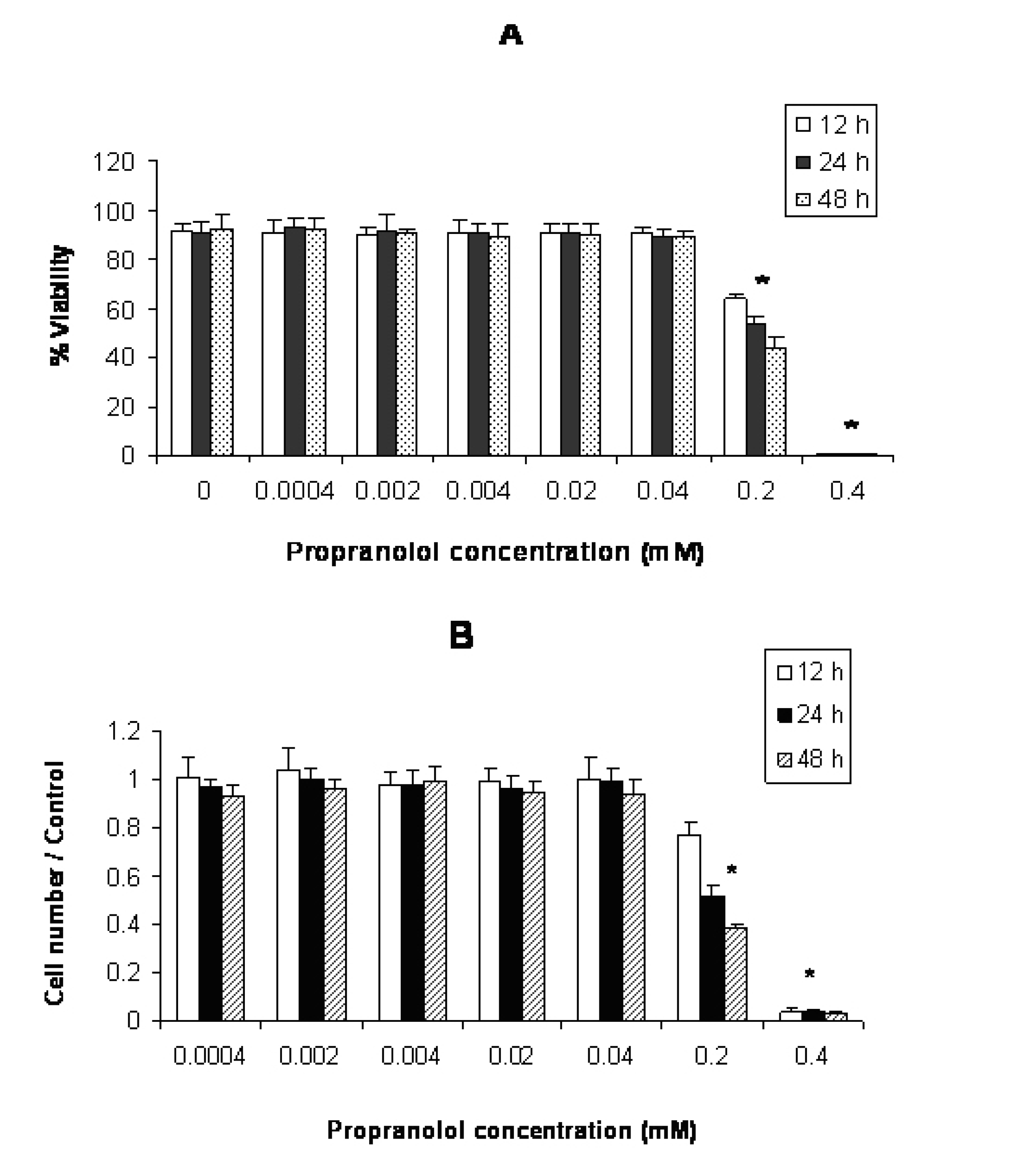
Figure 2. Effect of propranolol on proliferative responses of human leukemic Jurkat T-cell line. The Jurkat cells were treated with different concentrations of propranolol (0.0004 - 0.4 mM) for 12, 24 and 48 hours. The results are presented as % of viability demonstrated by trypan blue dye exclusion (TB) test (A) and cell number/control demonstrated by MTT assay (B). Data are mean ± SD of triplicate cultures. n = 3; P < 0.05 was considered significant.
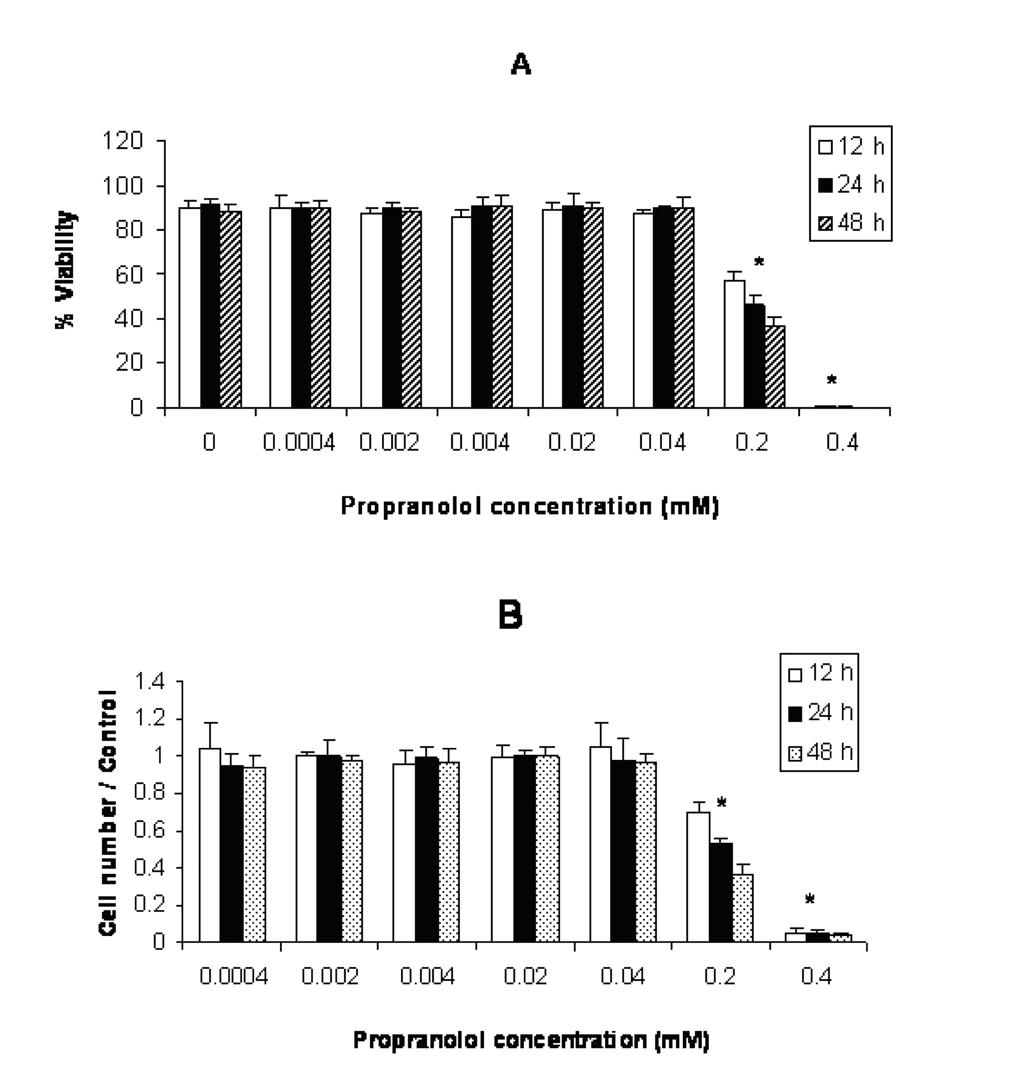
Figure 3. Effect of propranolol on proliferative responses of human leukemic U937 cell line. The U937 cells were treated with different concentrations of propranolol (0.0004 - 0.4 mM) for 12, 24 and 48 hours. The results are presented as % of viability demonstrated by trypan blue dye exclusion (TB) test (A) and cell number/control demonstrated by MTT assay (B). Data are mean ± SD of triplicate cultures. n = 3; P < 0.05 was considered significant.