Figures
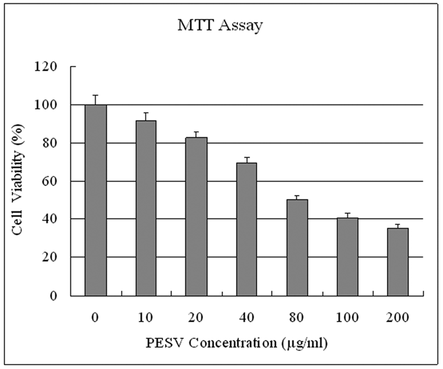
Figure 1. Effect of PESV on the growth of prostate cancer cells DU145. Cells were treated with PESV (10, 20, 40, 80, 100, and 200mg/L), and the percentage inhibition of cell growth was determined by MTT assay in a 96-well microtiter plate as detailed in Materials and Methods. Columns, mean of three separate experiments wherein each treatment was repeated in 10 wells; bars, SE.
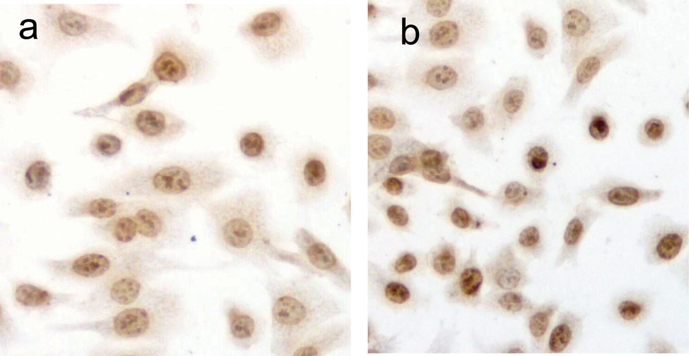
Figure 2. Detection of PESV-induced apoptosis on DU145 cells by DNA fragment TUNEL staining. Apoptotic nuclei and fragmented DNA were stained dark brown in treated cells (2b) but not in untreated controls (2a) in DU145 cells. Original magnification x400.
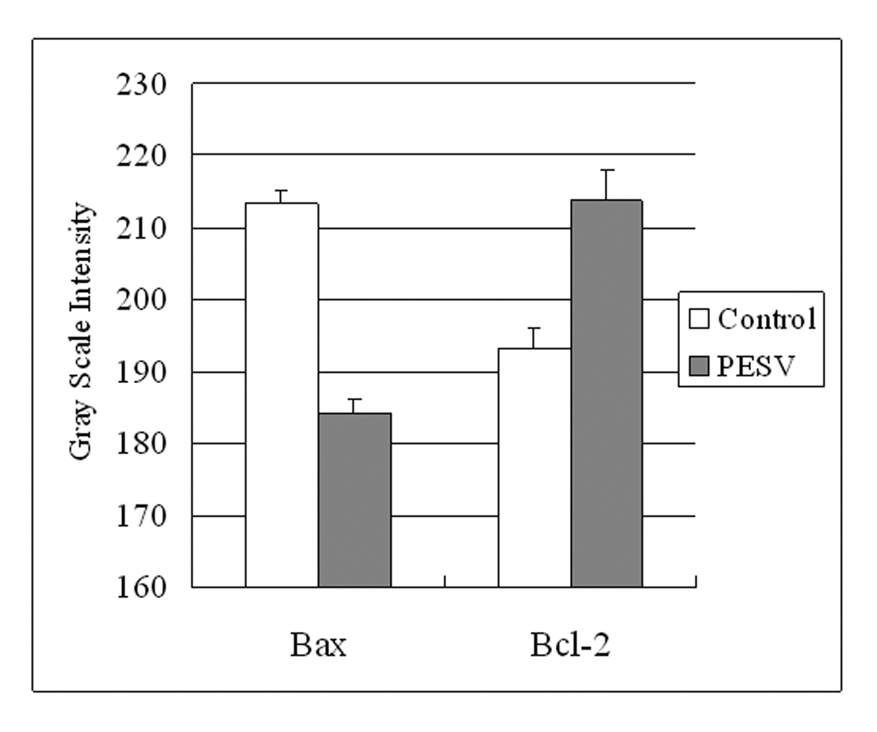
Figure 3. Grey Scale Intensity variants evaluated by Leica QWin V3 software of Bax and Bcl-2 immunoreactivity in DU145 cells treated with PESV and control group. Higher grey scale intensity standing for weaker protein expression, and lower, sronger protein expression. Columns, mean of three separate experiments conducted in triplicate with DU145 cells; bars, SE.
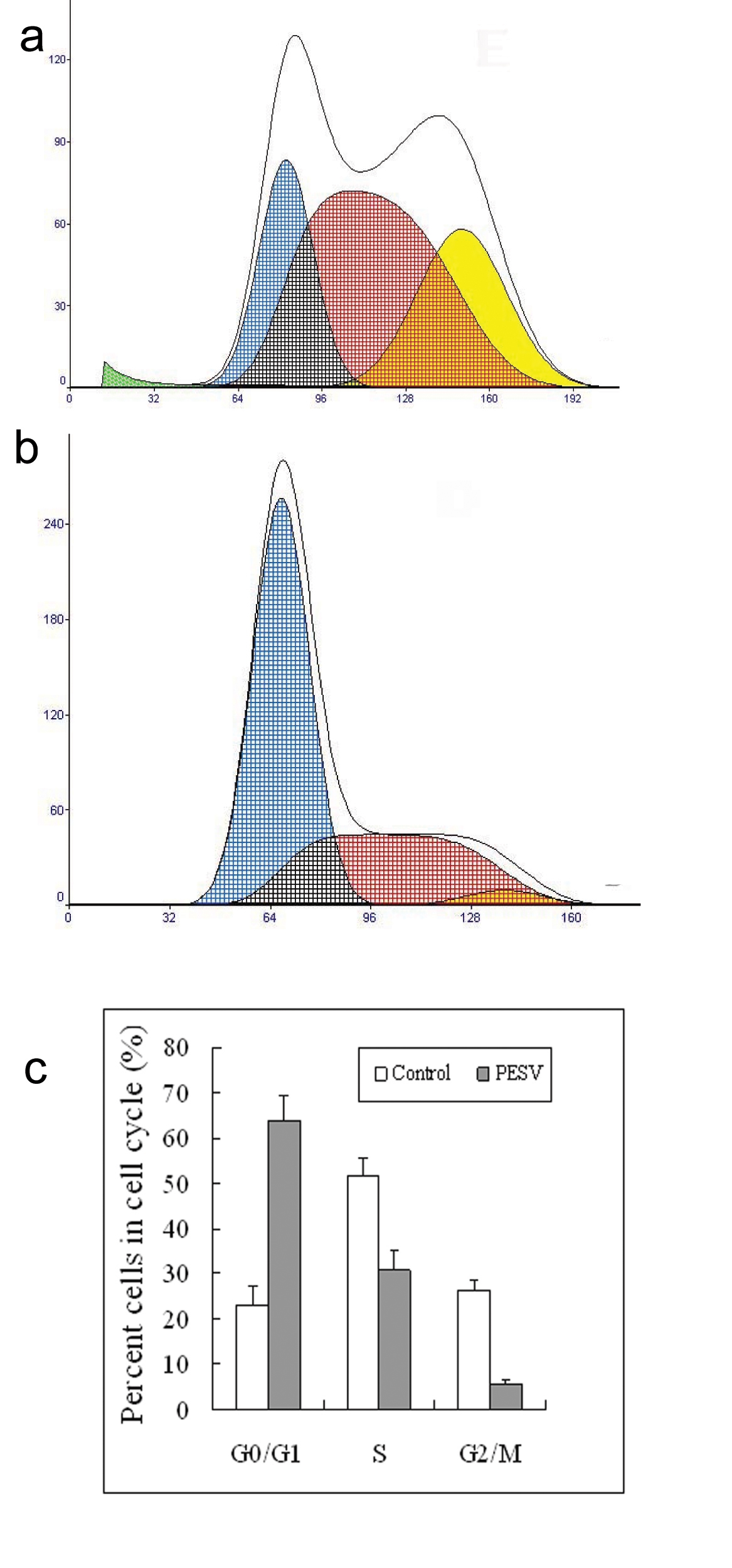
Figure 4. Representative DNA histograms and percentage of cells in different cell cycle phases after incubated with 40mg/L of PESV (B) for 48 h in human DU145 cells by flow cytometry. The growing cells (60% confluent) were treated with PESV (40mg/L) for 48 hours, and the DNA cell cycle analysis was done as described in Materials and Methods. Columns, mean of three separate experiments conducted in triplicate with DU145 cells; bars, SE.
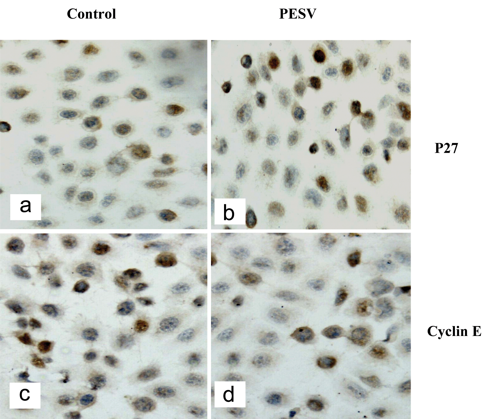
Figure 5. Representative image of cell cycle-related protein expression. P27kip1 (5a, 5b) and Cyclin E (5c, 5d). p27 expressed in the nuclei in DU145 cells treated with PESV (5b) is stonger than the control (5a); But Cyclin E expressed in the nuclei in DU145 cells treated with PESV (5d) is weaker than the control (5c).
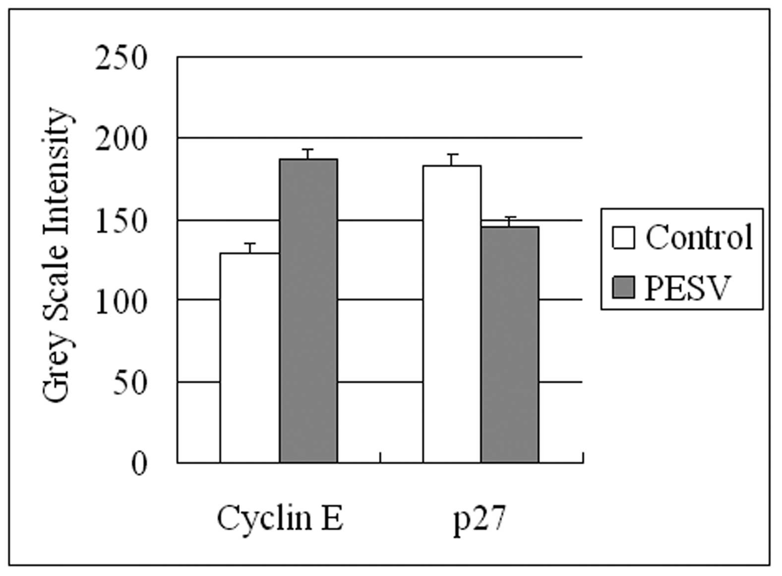
Figure 6. Grey Scale Intensity variants evaluated by Leica QWin V3 software of Cyclin E and p27 immunoreactivity in DU145 cells treated with PESV and control group. Higher grey scale intensity standing for weaker protein expression, and lower, sronger protein expression.
Table
Table 1. Effect of PESV on apoptosis in prostate cancer cells DU145 (TUNEL Assay)
| Treatment | Apoptosis index (%) | P value |
|---|
| Control | 3.6 ± 0.02 | |
| PESV (40 mg/L) | 8.3 ± 0.04 | 0.001 |