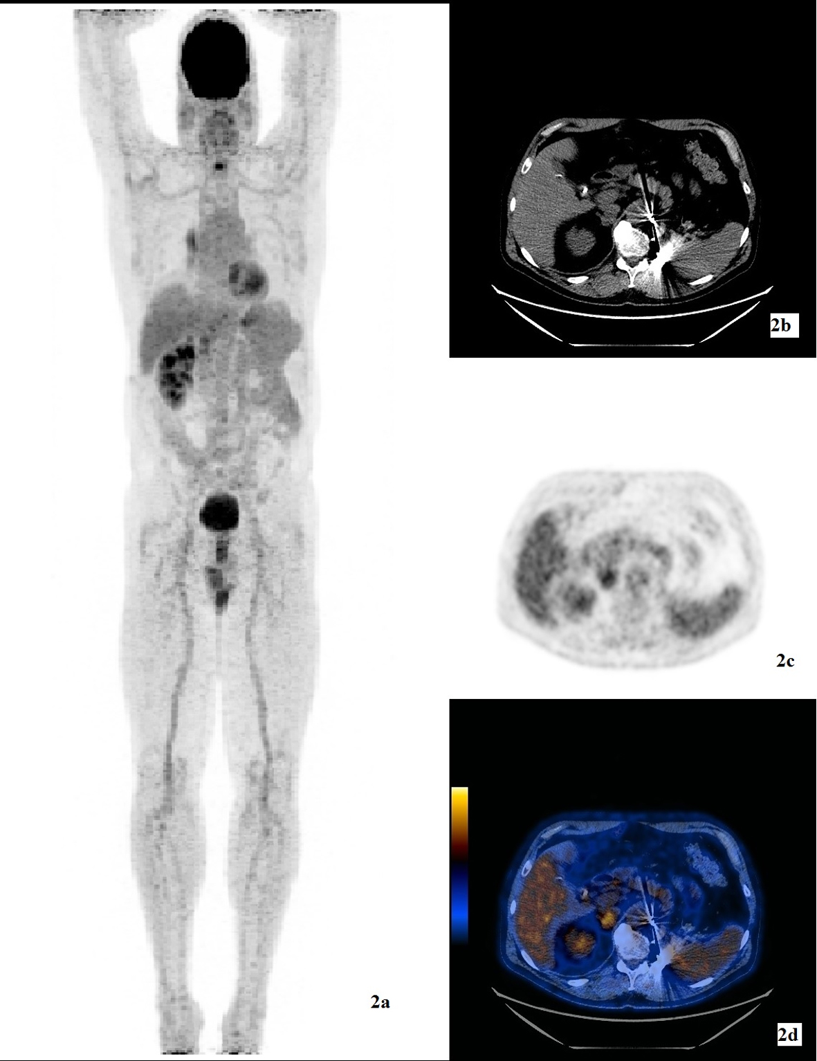
Figure 1. (a) 3D MIP image of FDG-PET/CT study showing a hypermetabolic lesion (arrow) in the gallbladder. (b) CT showing a non-specific hyperdense nodule within the gallbladder. The gallbladder was otherwise normal with no wall thickening and no pericholecystic fluid. (c) FDG emission image. (d) Hybrid FDG-PET/CT image showing a hypermetabolic soft tissue density of SUVmax 6 (arrow) within the fundus of the gallbladder.