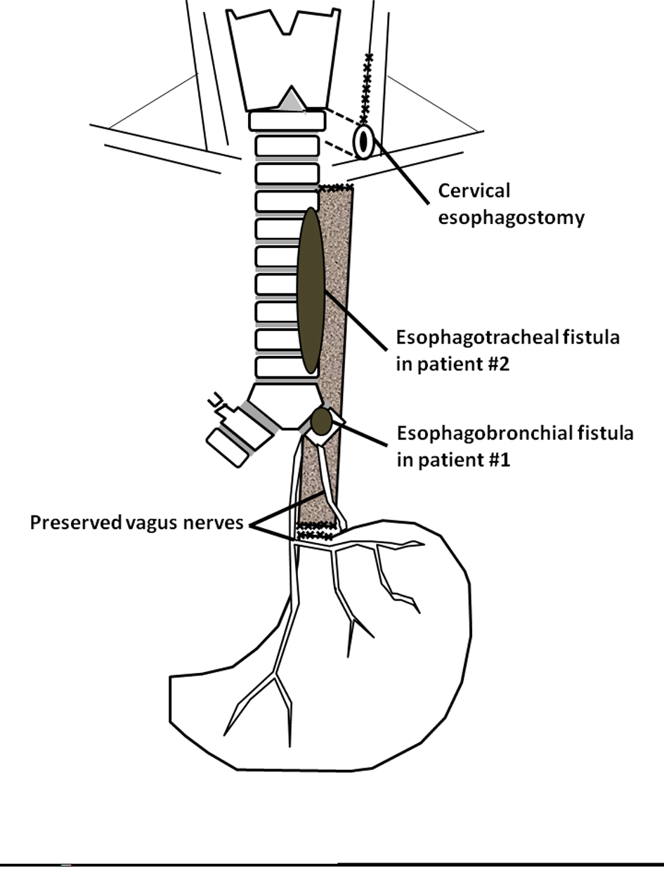
Figure 1. Surgical technique of bipolar esophagus exclusion (modified from Maillard et al [5]).
| Journal of Clinical Medicine Research, ISSN 1918-3003 print, 1918-3011 online, Open Access |
| Article copyright, the authors; Journal compilation copyright, J Clin Med Res and Elmer Press Inc |
| Journal website http://www.jocmr.org |
Case Report
Volume 5, Number 2, April 2013, pages 140-143
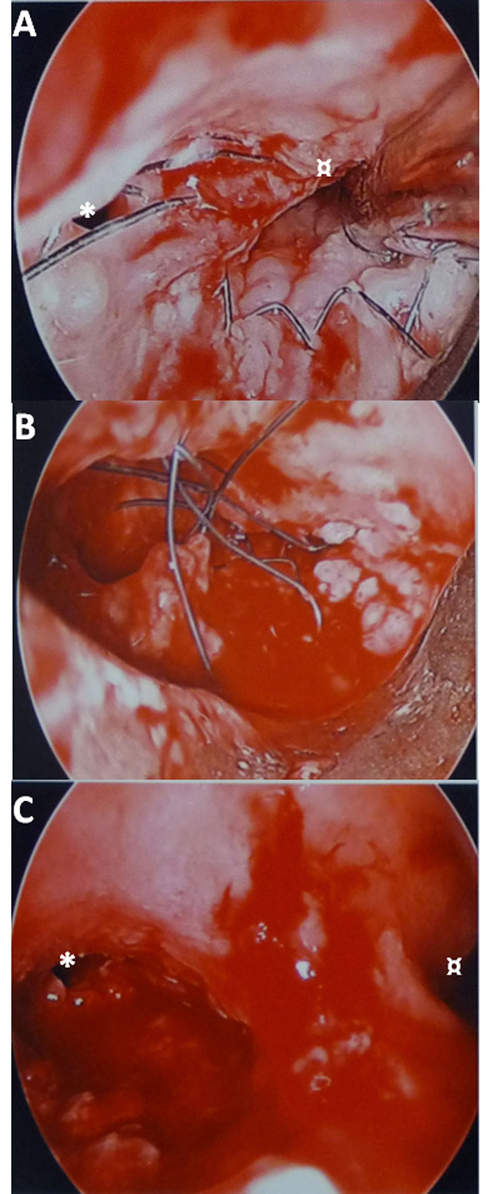
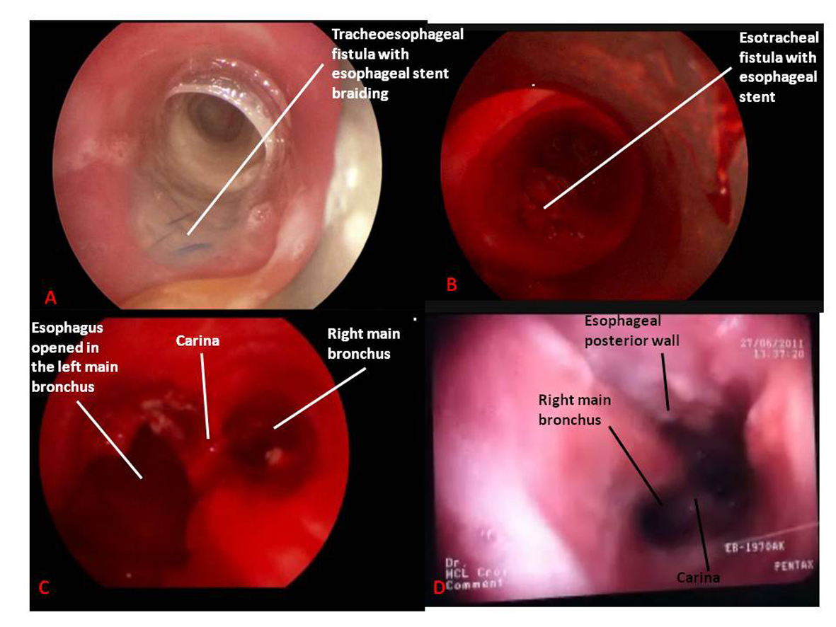
Transtracheal Esophageal Stent Removal: A Case-Series
Figures