Journal of Clinical Medicine Research, ISSN 1918-3003 print, 1918-3011 online, Open Access
Article copyright, the authors; Journal compilation copyright, J Clin Med Res and Elmer Press Inc
Journal website http://www.jocmr.org

Original Article

Volume 5, Number 2, April 2013, pages 127-131


Terazosin Treatment Induces Caspase-3 Expression in the Rat Ventral Prostate

Figure

Figure 1.
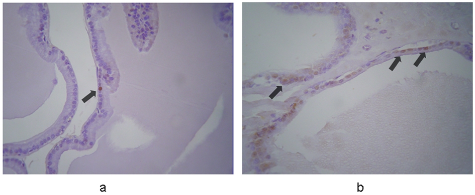
Figure 1. Immunohistochemical expression of caspase-3 in the prostates from (a) control and (b) terazosin-treated rats (magnification × 100). In controls caspase-3 is expressed in epithelial cells weakly and sporadically. In contrast, in terazosin-treated specimens, stronger expression is evident in a significantly larger proportion of epithelial cells, mainly in those detached to the lumen of the acini. The arrows show positive nuclei in caspase-3 expression.

Table

Table 1. Body Weight (BW), Absolute Prostate Weight (APW), and Relative Prostate Weight (RPW) of Terazosin-Treated (Group A) and Control (Group B) Rats (Mean ± Standard Deviation)
 
BW (g)APW (g)RPW (mg/g)
startend
Group A430 ± 39459 ± 420.61 ± 0.111.28 ± 0.29
Group B418 ± 19490 ± 270.52 ± 0.021.07 ± 0.05