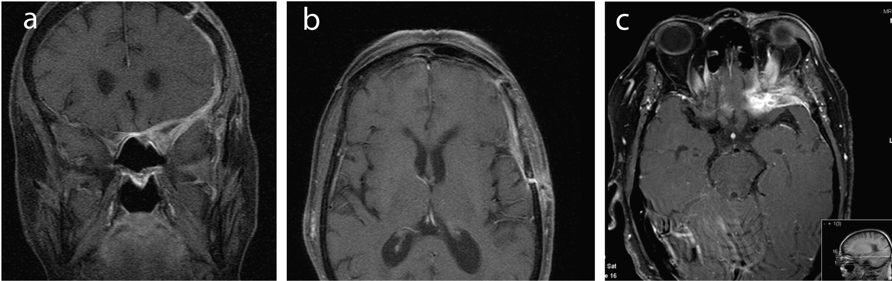
Figure 1. (a-c) MRI images (coronal and horizontal cuts) showing a pachymeningeal thickening around the left anterior clinoid process and lesser wing of the sphenoid, that extends to the left anterior cranial fossa and the anterior part of the middle cranial fossa; it also extends to the orbitary apex through the left optic foramen and superior orbital fissure, affecting the extraocular orbitary muscles, compressing the optic nerve and infiltrating the bone marrow of the lesser wing of the sphenoid and the orbitary wall. This lesion was hypointense on both T1- and T2-weighted images and proeminently enhanced after administration of gadolinium.