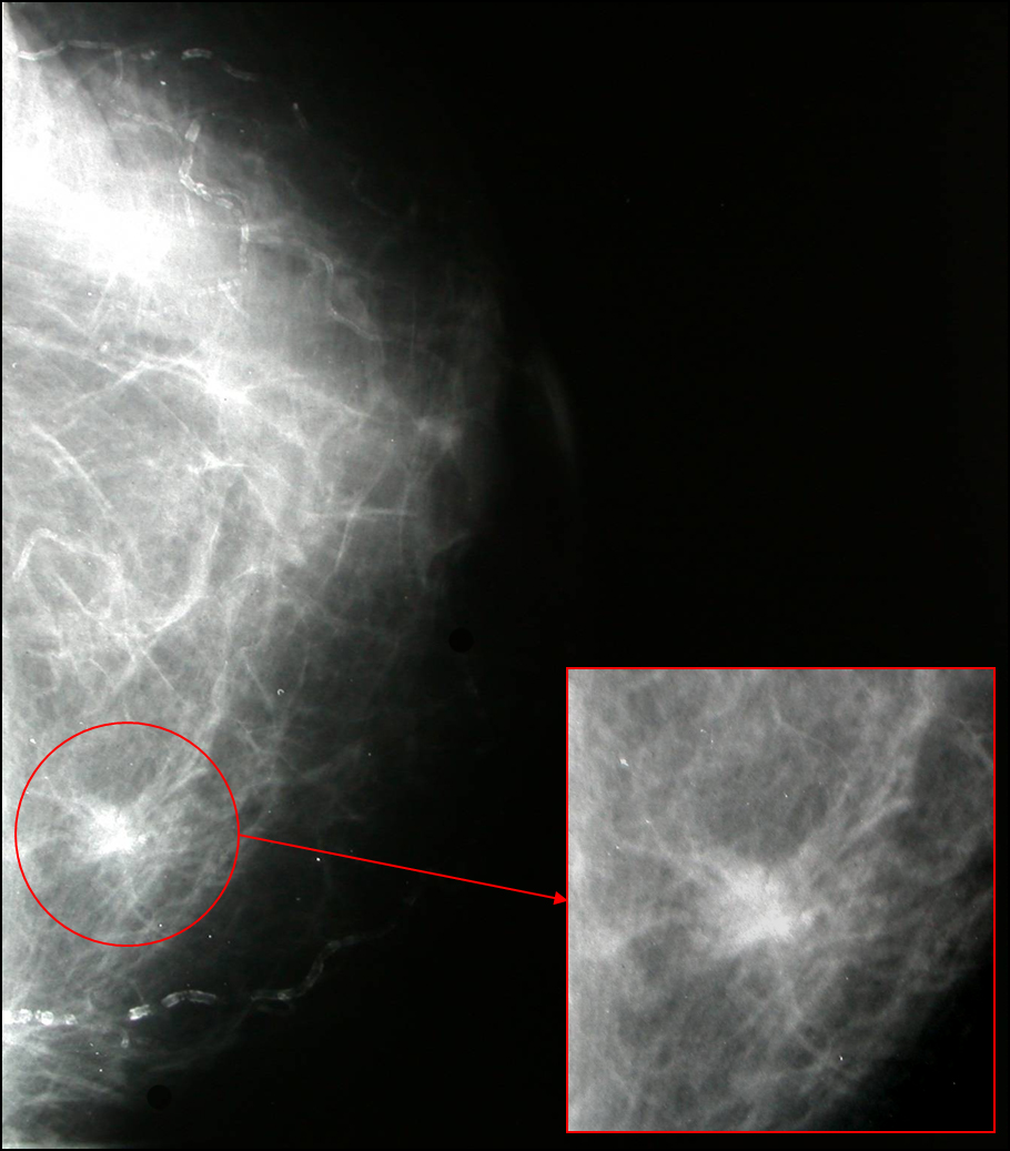
Figure 1. Mammography showed an ill-defined, high-density spiculated mass.
| Journal of Clinical Medicine Research, ISSN 1918-3003 print, 1918-3011 online, Open Access |
| Article copyright, the authors; Journal compilation copyright, J Clin Med Res and Elmer Press Inc |
| Journal website http://www.jocmr.org |
Case Report
Volume 2, Number 4, August 2010, pages 185-188
Cutaneous Granular Cell Tumor of the Breast: A Clinical Diagnostic Pitfall
Figures