Journal of Clinical Medicine Research, ISSN 1918-3003 print, 1918-3011 online, Open Access
Article copyright, the authors; Journal compilation copyright, J Clin Med Res and Elmer Press Inc
Journal website http://www.jocmr.org

Case Report

Volume 2, Number 4, August 2010, pages 194-197


Wilms’ Tumor in a 37-Year-Old

Figures

Figure 1.
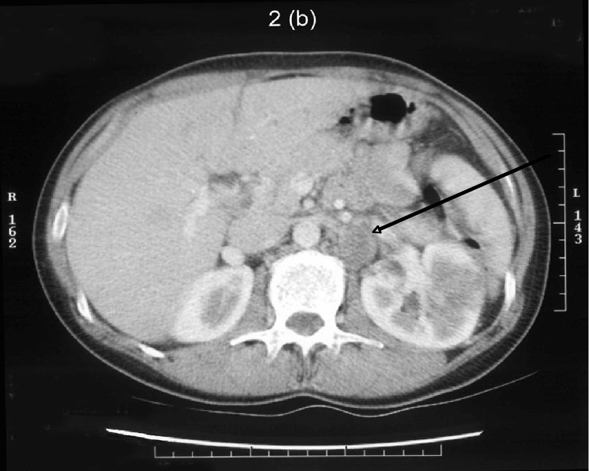
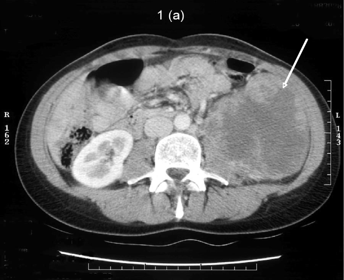
Figure 1. Post contrast CT demonstrating a 10 cm heterogeneously enhancing mass arising from the lower pole of the left kidney. It is centrally necrotic. The mass encroaches onto the left psoas and blocks the fat plane between itself and the left psoas muscle.
Figure 2.
Figure 2. A 2.5 cm pathologically enhanced lymph node crossed by the left renal vein.
Figure 3.
Figure 3. Absent function in inferior pole of the left kidney
Figure 4.
Figure 4. A triphasic adult Wilms’ tumor exhibiting a epithelial component (A), stromal component (B) and small areas of blastema (C)
Figure 5.
Figure 5. WT-1 staining showing strong nuclear positivity in the glandular component and weak nuclear positivity in the stromal component