Journal of Clinical Medicine Research, ISSN 1918-3003 print, 1918-3011 online, Open Access
Article copyright, the authors; Journal compilation copyright, J Clin Med Res and Elmer Press Inc
Journal website http://www.jocmr.org

Case Report

Volume 10, Number 8, August 2018, pages 665-667


Unusual Features of Clear Cell Adenocarcinoma of the Female Urethra: A Case Report and Literature Review

Figures

Figure 1.
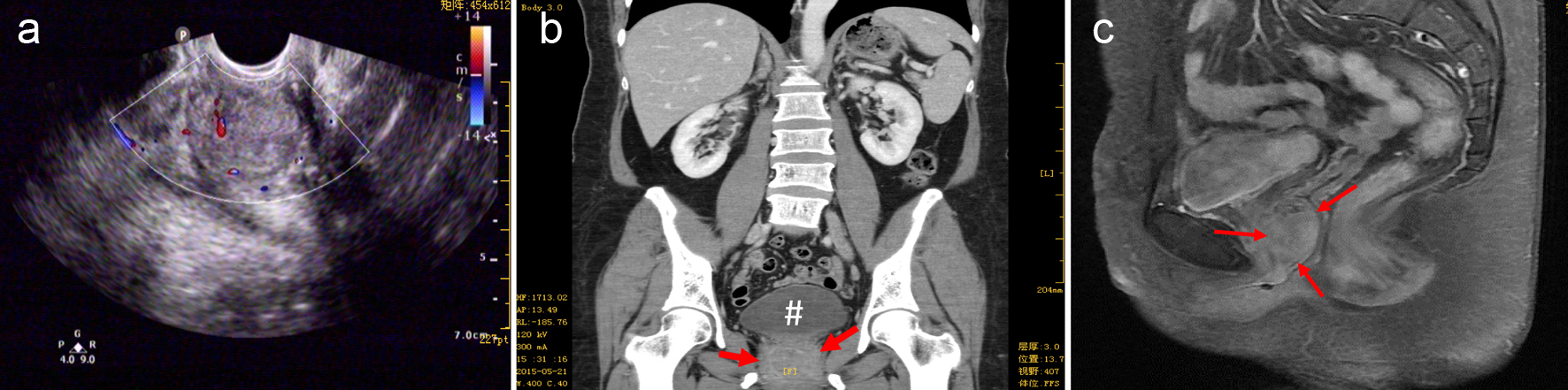
Figure 1. Imaging findings. (a) A hypoechoic mass (about 31 × 26 mm) with well-defined margins between the anterior vaginal wall and the posterior urethral wall was seen by the ultrasound exam. (b) CT scan (feet first-supine position) demonstrated the mass (indicated by the red arrows) was inferior to the urinary bladder (oval-shaped as indicated by #) approximately the level of vaginal fornix and had smooth margins; (c) the sagittal magnetic resonance T1-weighted imaging showed the tumor (indicated by the red arrows) located at the anterior vaginal wall.
Figure 2.
Figure 2. Histology and immunohistochemistry exams. Panel a is a representative image of HE staining that shows numerous tumor cells with clear cytoplasm, a key feature of CCAU. Hobnail-shaped cells and basally located pleomorphic nuclei without prominent nucleoli were also seen. Panel b shows strong staining for CK7 antigen by immunohistochemical analysis.