Journal of Clinical Medicine Research, ISSN 1918-3003 print, 1918-3011 online, Open Access
Article copyright, the authors; Journal compilation copyright, J Clin Med Res and Elmer Press Inc
Journal website http://www.jocmr.org

Case Report

Volume 10, Number 6, June 2018, pages 527-530


Dissecting Vertebral Artery Aneurysm Presenting Regrowth After Stent-Assisted Coil Embolization in Acute Stage

Figures

Figure 1.
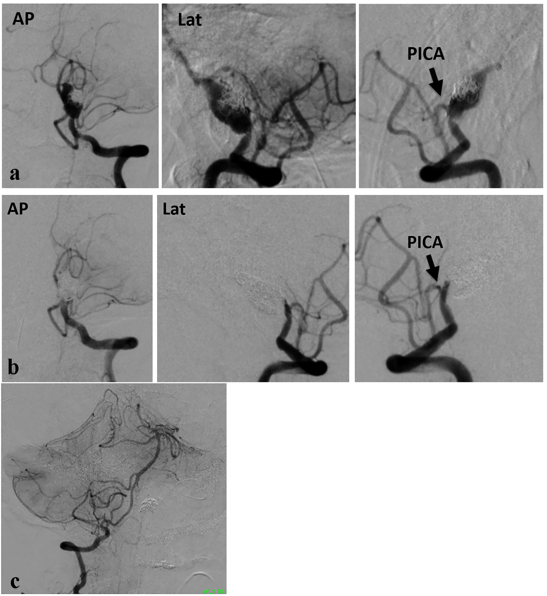
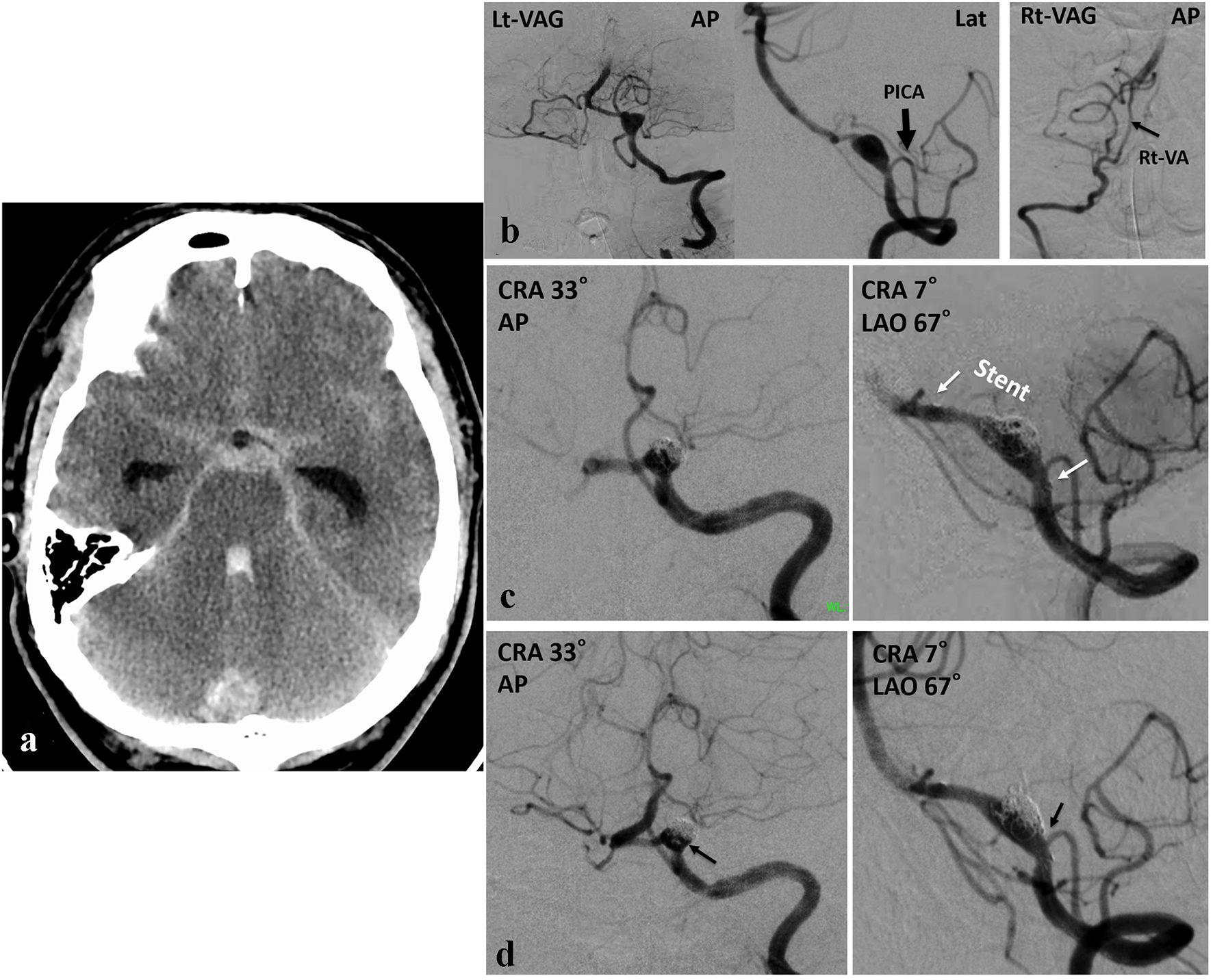
Figure 1. (a) CT demonstrating SAH. (b) Angiography demonstrating DVAA on the left VA. The PICA originating proximal to the dissected portion. The diameter of the affected left VA is thicker than that of the right. (c) Angiography showing two working angles. A stent is placed in the left VA, covering from distal normal VA to proximal to the PICA origin including dissection. (d) Post-embolization angiography showing occlusion of the dissected portion and patency of the left VA. The residual cavity besides the VA and a small portion at the distal to the PICA origin are opacified (arrow).
Figure 2.
Figure 2. (a) Angiography performed on the 10th day showing recurrence of DVAA. Dissection was extended identical to the location of stent. (b) Post-embolization angiography showing complete occlusion of the left VA including aneurysm. A small part of the VA remains distal to the PICA. (c) The right VA has a significant diameter.