Journal of Clinical Medicine Research, ISSN 1918-3003 print, 1918-3011 online, Open Access
Article copyright, the authors; Journal compilation copyright, J Clin Med Res and Elmer Press Inc
Journal website http://www.jocmr.org

Case Report

Volume 8, Number 5, May 2016, pages 420-423


A Very Rare Cause of Anal Atresia: Currarino Syndrome

Figures

Figure 1.
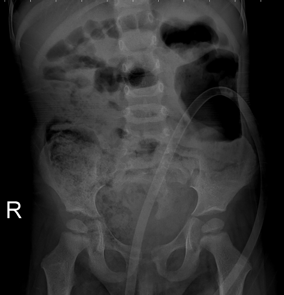
Figure 1. An abdominal radiograph: a defect at the right lower side of the sacrum, and a dysplasic image displaying deviation to the left (scimitar sacrum).
Figure 2.
Figure 2. An ultrasound examination revealed a multilocular septated cystic lesion measuring 5.7 × 6 cm in the presacral area.
Figure 3.
Figure 3. Abdominal tomogram: hypoplasic appearance of the right sides of S3, and vertebras inferior to S3, leftward deviation of sacrum (scimitar sacrum), and a presacral cystic lesion.
Figure 4.
Figure 4. Lumbosacral MRI: widening of the right S3-S4 and S4-S5 neural foramens, and a multilocular, septated anterior sacral meningocele (ASM) which continued with sacral spinal canal anteriorly displaced bladder, and rectum were observed.