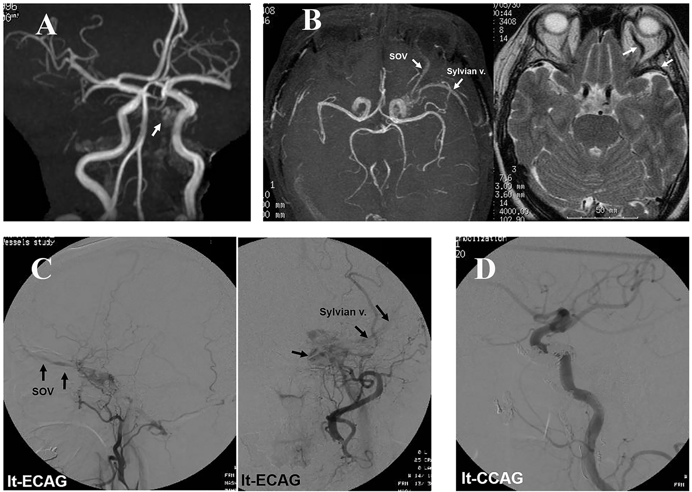
Figure 1. Case 1: (A) MRA obtained 3 months after the initial headache showing an abnormal intensity in the left CS (arrow), but no dilatation of the SOV. (B) MRI demonstrating blood reflux to the left CS, SOV, and Sylvian vein after the patient had become aware of eye symptoms at the eighth month. (C) Angiography showing CS-dAVF reflux to the left SOV and Sylvian vein. (D) Post-embolization angiography showing no blood reflux to the CS.
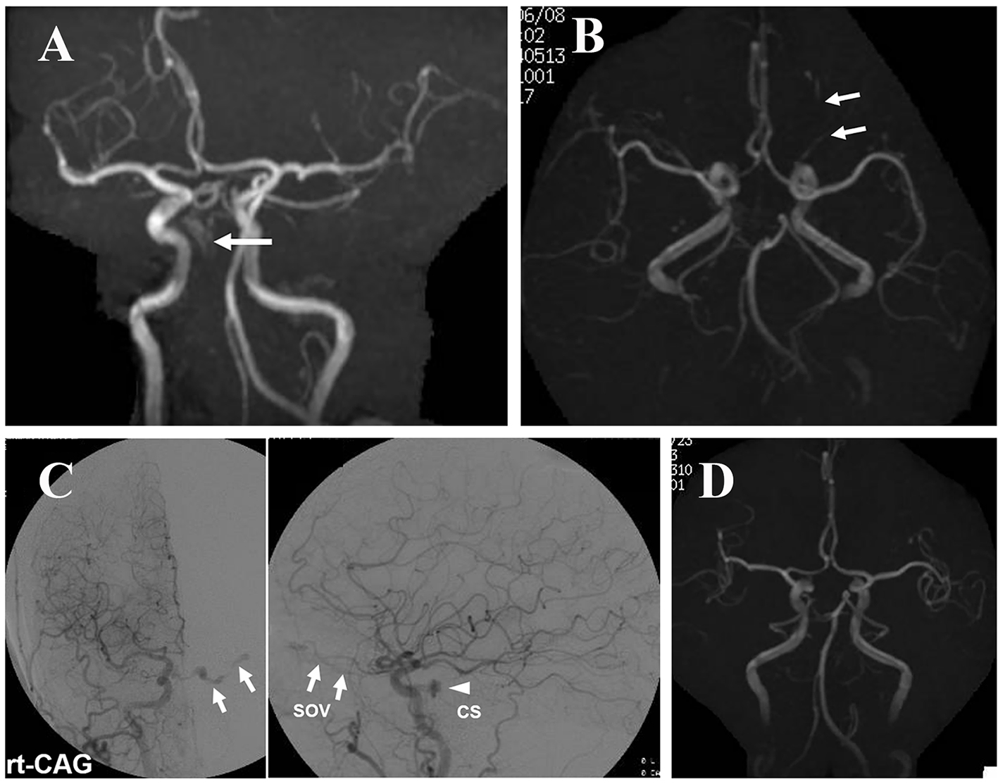
Figure 2. Case 2: (A) MRA performed 1 month after the initial headache demonstrating an abnormal intensity in the right CS (arrow). Reflux to and dilatation of the SOV are not apparent. (B) MRI after the patient had become aware of double vision, again showing blood reflux to and dilatation of the left SOV (arrows). (C) Angiography showing arteriovenous shunt to the right CS and reflux to the left SOV. No blood reflux to a cortical vein is observed. (D) MRA after manual compression of the carotid arteries showing the disappearance of blood reflux to the left SOV.
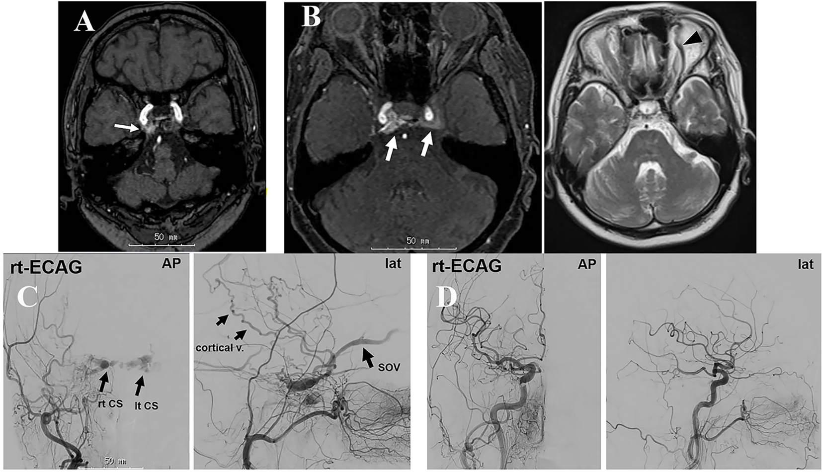
Figure 3. Case 3: (A) MRI obtained 2 months after the initial pain demonstrating an abnormal intensity in the right CS (arrow). (B) MRI/MRA obtained 10 months after onset showing abnormal blood flow in the bilateral CS (arrows) and dilatation of the left SOV (arrowhead). (C) Angiography showing abnormal blood flow in the right CS and SOV. The refluxed blood flows to the left CS via the intercavernous sinus and further to the left SOV. (D) Post-embolization angiography showing the disappearance of the abnormal blood flow.