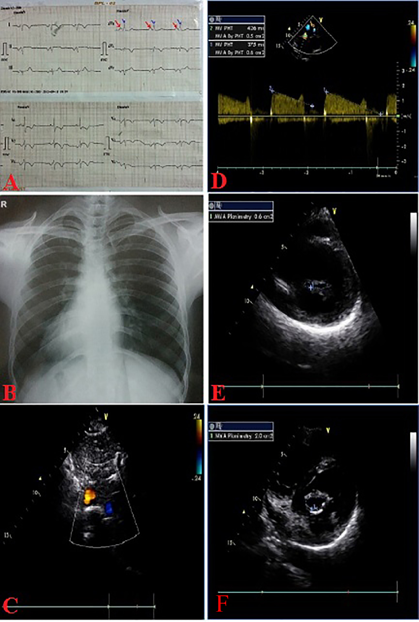
Figure 1. ECG (A); chest X-ray (B) PA view showing situs inversus totalis; subcostal echo showing situs inversus (C), critical MS by PHT (D), planimetry (E), post-PTMC MVA-2.0 cm2 (F).
| Journal of Clinical Medicine Research, ISSN 1918-3003 print, 1918-3011 online, Open Access |
| Article copyright, the authors; Journal compilation copyright, J Clin Med Res and Elmer Press Inc |
| Journal website http://www.jocmr.org |
Case Report
Volume 8, Number 4, April 2016, pages 351-355
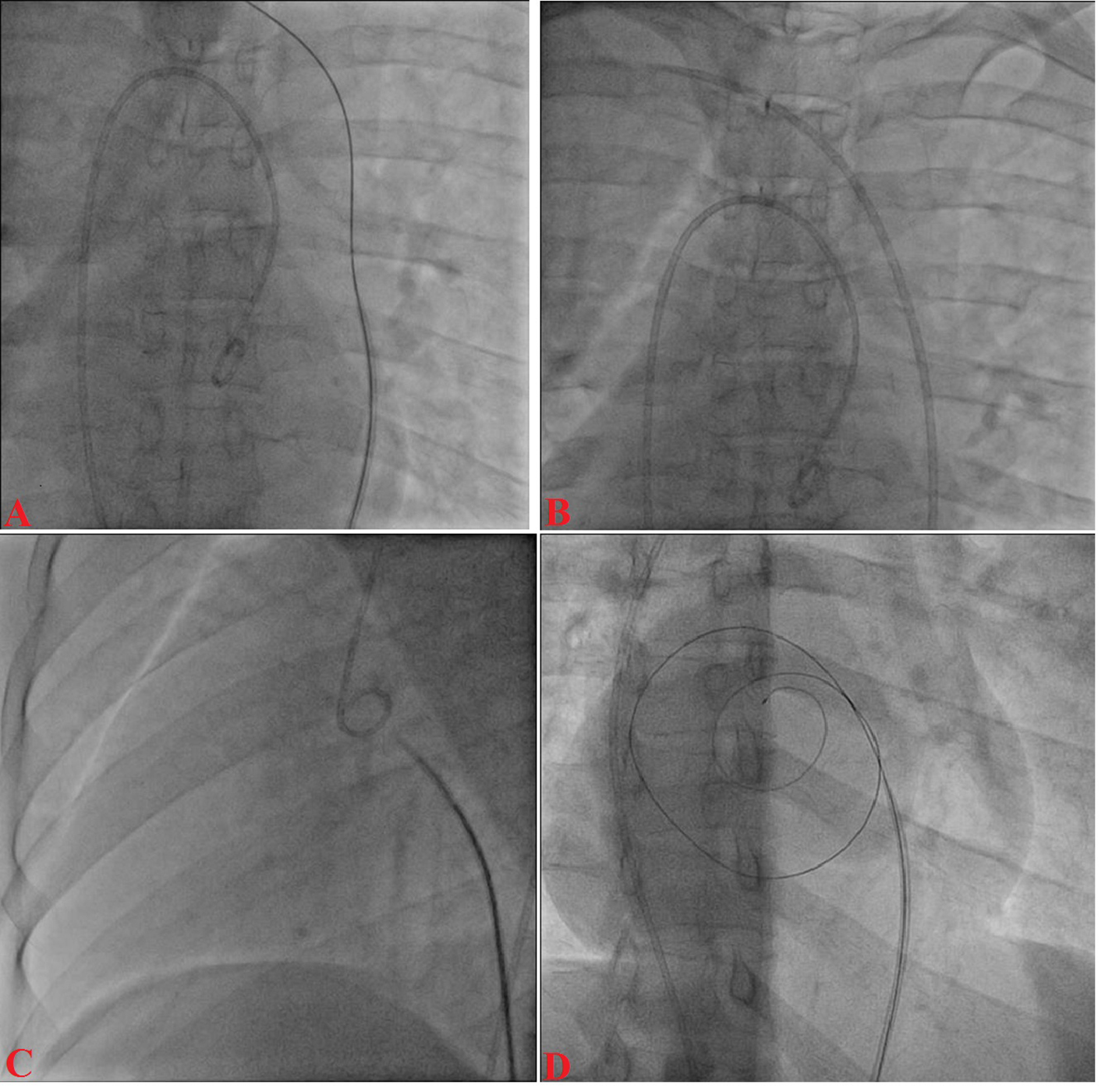
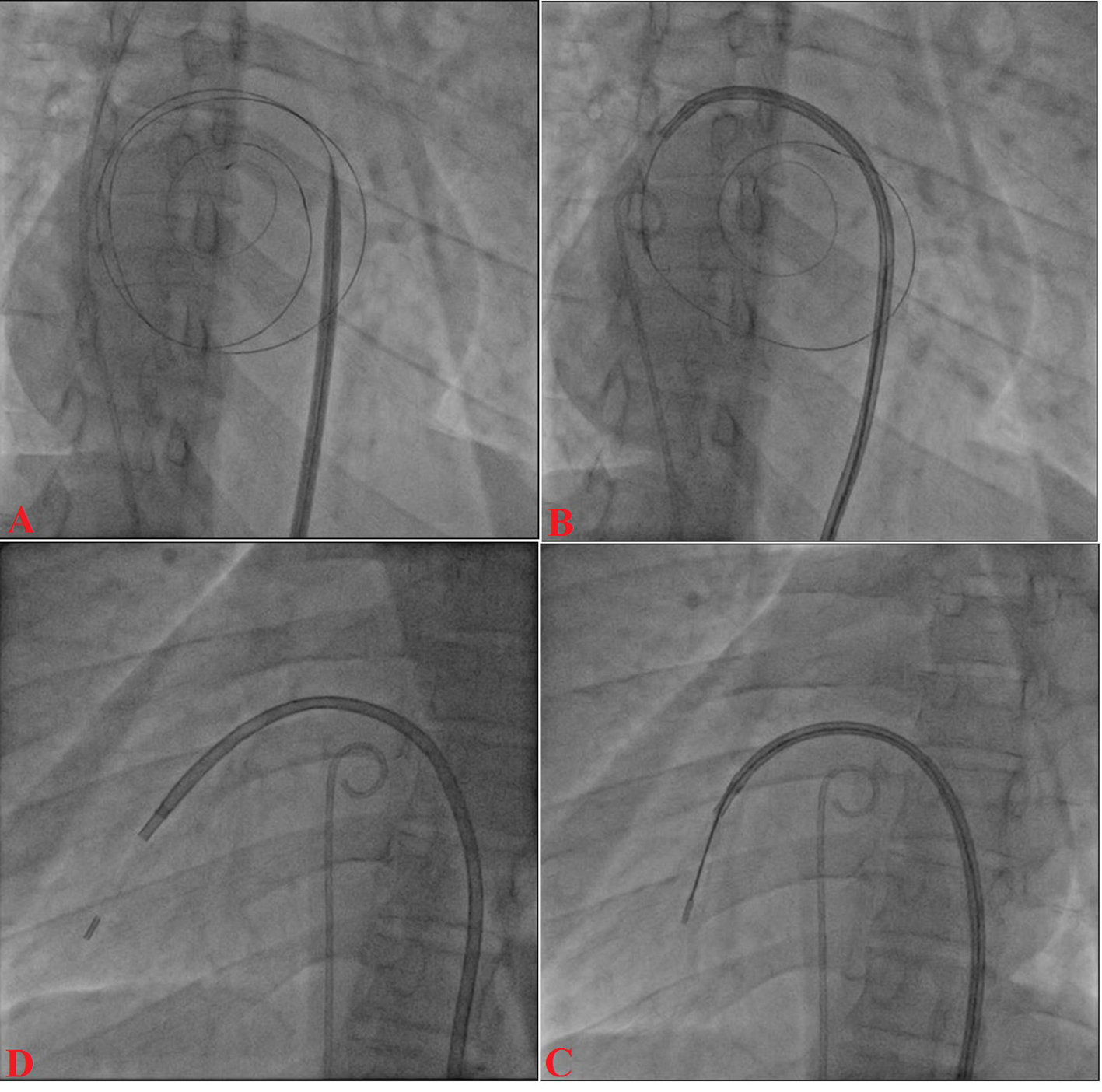
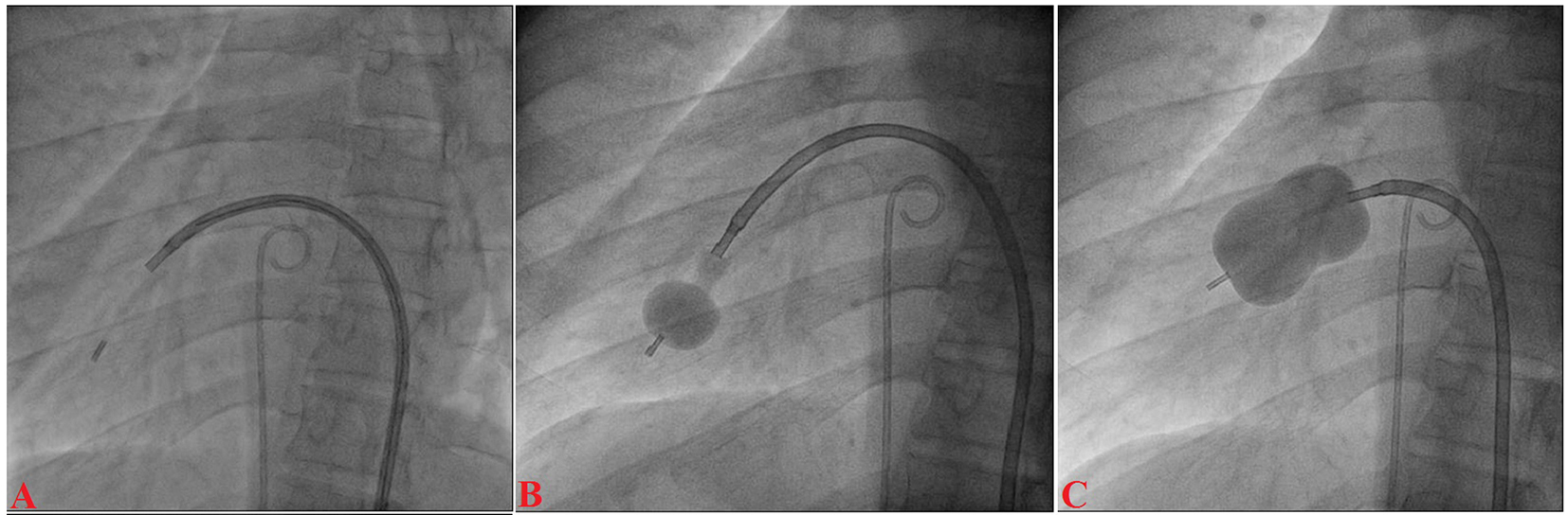
Percutaneous Mitral Valvotomy in a Case of Situs Inversus Totalis and Juvenile Rheumatic Critical Mitral Stenosis: Case Report
Figures
Table
| Steps | Conventional PTMC | PTMC in dextrocardia |
|---|---|---|
| Trans-septal catheterization | Right groin | Left groin |
| Descent of needle assembly | 4 - 6 o’clock position | 7 - 9 o’clock position |
| Septal puncture | AP view | Pseudo AP view |
| LAO view | RAO view | |
| Crossing of mitral valve and balloon dilatation | RAO view | LAO view |