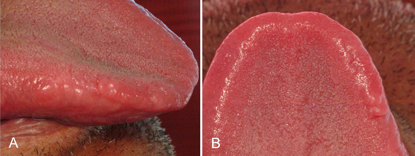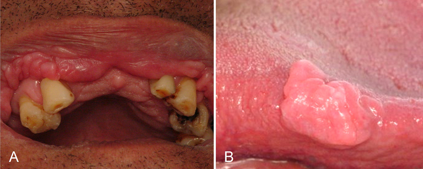
Figure 1. Multiple papules and nodules, sessile, coalescent, normal in colour, located on the upper lip mucosa and causing double lip appearance (A). Solitary tongue nodule, sessile, measuring approximately 20 mm on its largest diameter, with smooth and lobulated surface, similar in colour to the surrounding mucosa (B).
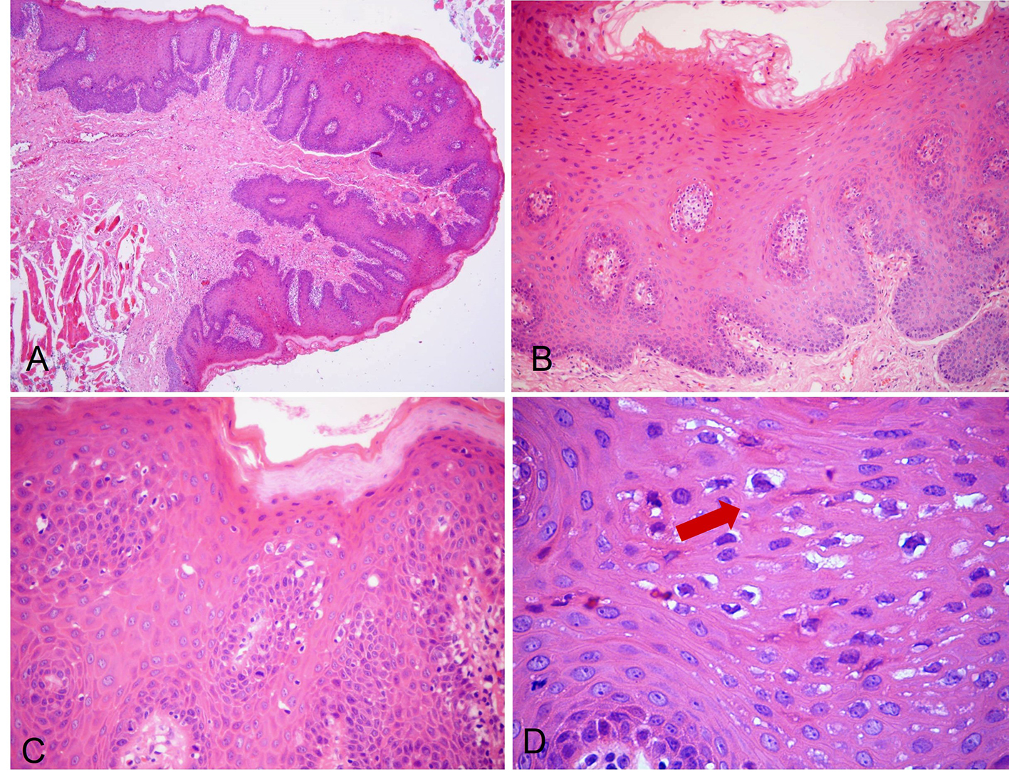
Figure 3. H&E-stained photomicrographs displaying characteristic features of FEH. Low-power magnification shows pseudocarcinomatous hyperplasia, marked acanthosis and elongated rete ridges (A, × 40; B, × 100). Spinous layer with parakeratosis, anisokaryosis and typical koilocytosis (C, × 200). High magnification allows clearer depiction of koilocytosis showing keratinocytes with pyknotic nuclei, surrounded by clears areas (D, × 400, arrow).