Journal of Clinical Medicine Research, ISSN 1918-3003 print, 1918-3011 online, Open Access
Article copyright, the authors; Journal compilation copyright, J Clin Med Res and Elmer Press Inc
Journal website http://www.jocmr.org

Case Report

Volume 8, Number 3, March 2016, pages 254-256


Histoplasma capsulatum: An Unusual Case of Pericardial Effusion and Coarctation of the Aorta

Figures

Figure 1.
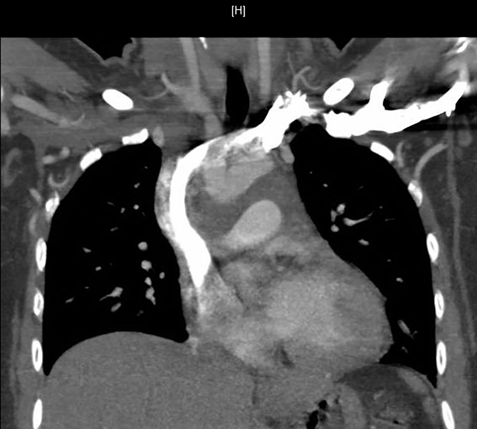
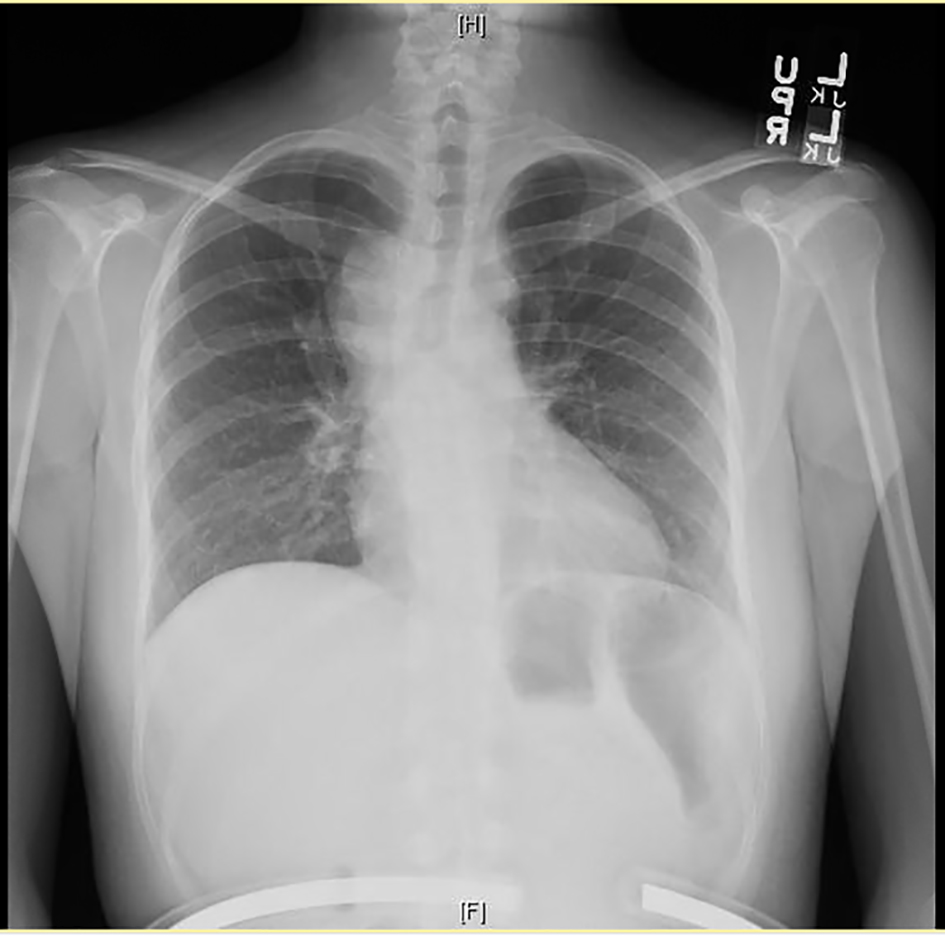
Figure 1. Chest X-ray showing right peri-hilar mass with tracheal compression.
Figure 2.
Figure 2. Computed tomography (CT) of chest showing mediastinal mass and mediastinal lymphadenopathy.
Figure 3.
Figure 3. Wedge resection of right middle lobe of lung showing coalescing fibrocaseous granulomas (hematoxylin-eosin stain, magnification, × 100).
Figure 4.
Figure 4. Wedge resection of right middle lobe of lung depicting giant cell histiocytes, epithelioid histiocytes, lymphocytes and fibrocaseous necrosis (hematoxylin-eosin stain, magnification, × 100).
Figure 5.
Figure 5. Wedge resection of right middle lobe of lung, magnification of fibrocaseous necrosis center of granuloma with budding yeast consistent with histoplasmosis species (Grocott methenamine silver stain, magnification, × 400).
Figure 6.
Figure 6. Computed tomography (CT) of chest showing pericardial effusion with pericardial thickening.