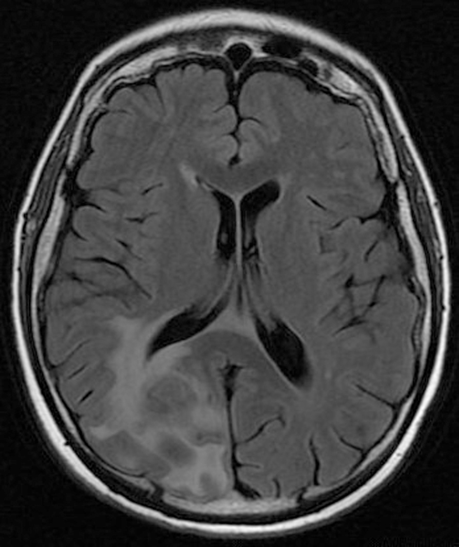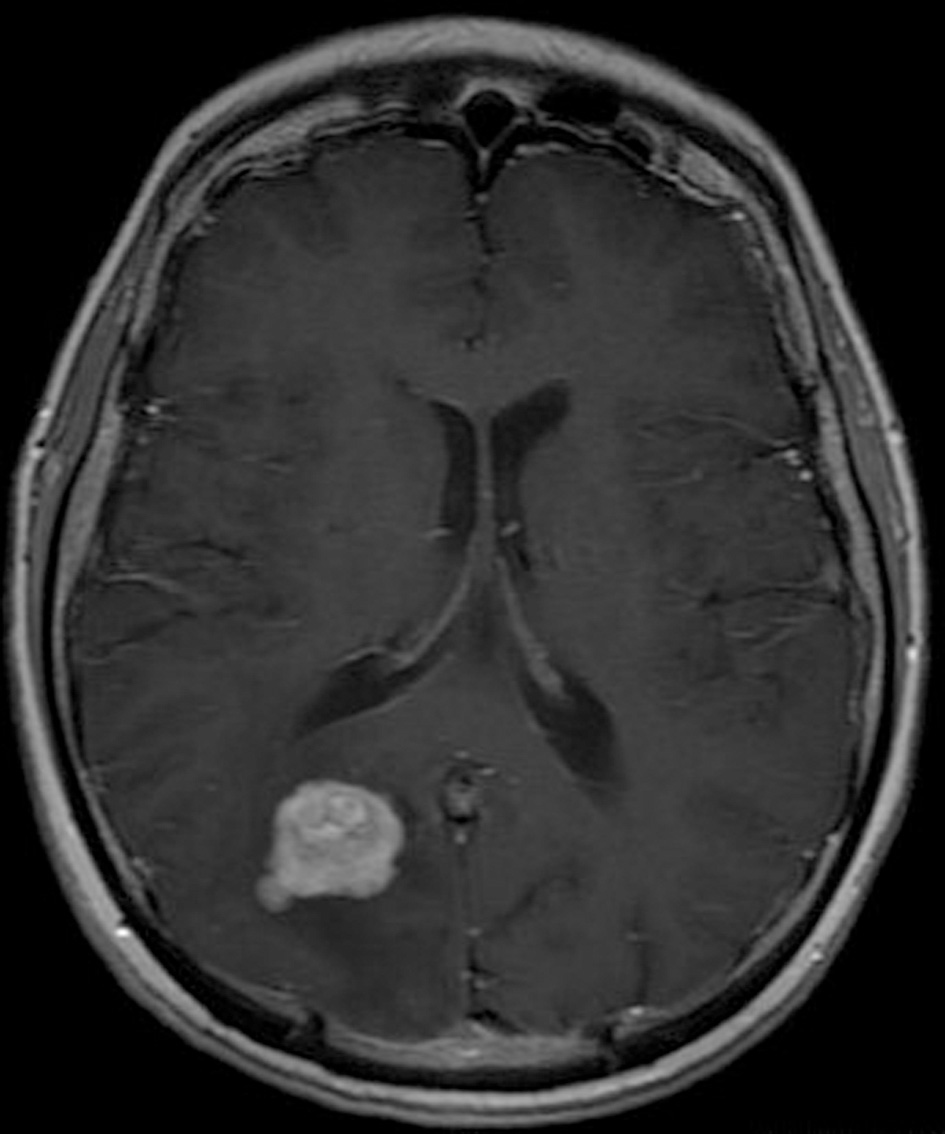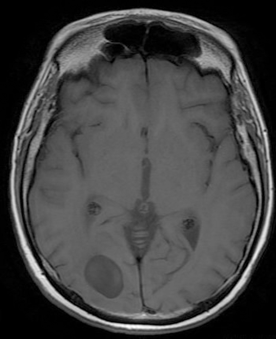
Figure 1. Pre-operative brain MRI T1 sequence.
| Journal of Clinical Medicine Research, ISSN 1918-3003 print, 1918-3011 online, Open Access |
| Article copyright, the authors; Journal compilation copyright, J Clin Med Res and Elmer Press Inc |
| Journal website http://www.jocmr.org |
Case Report
Volume 7, Number 12, December 2015, pages 1007-1012
Primary Diffuse Large B-Cell Lymphoma of Central Nervous System: Is Still Surgery an Unorthodox Treatment?
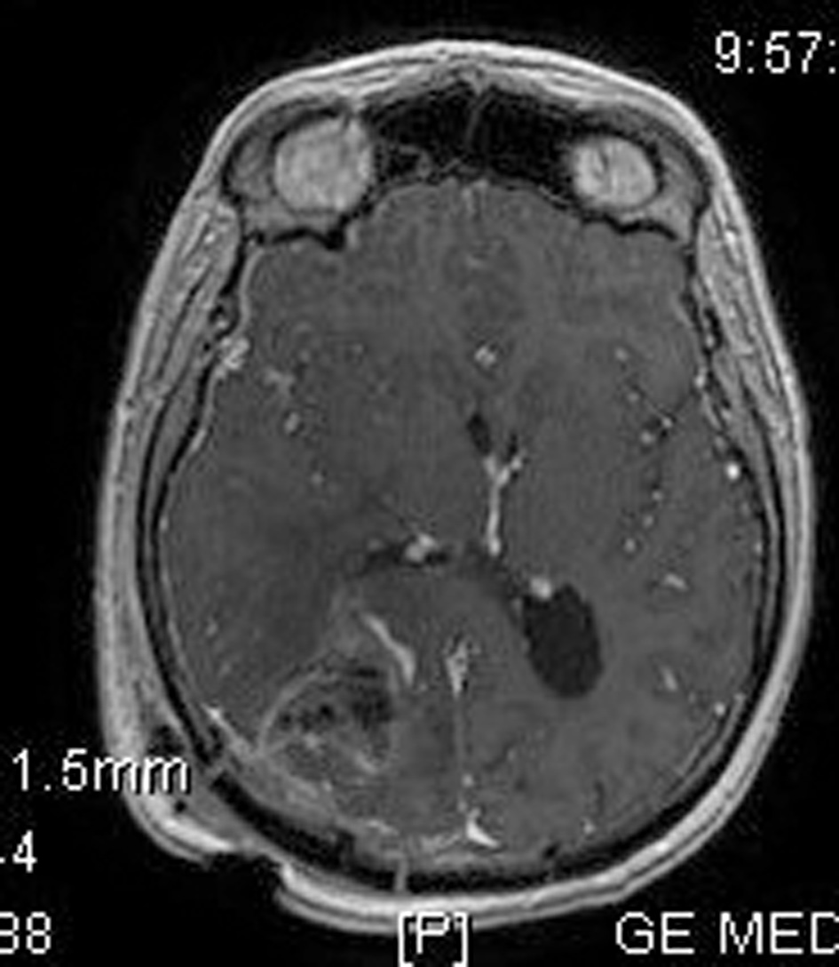
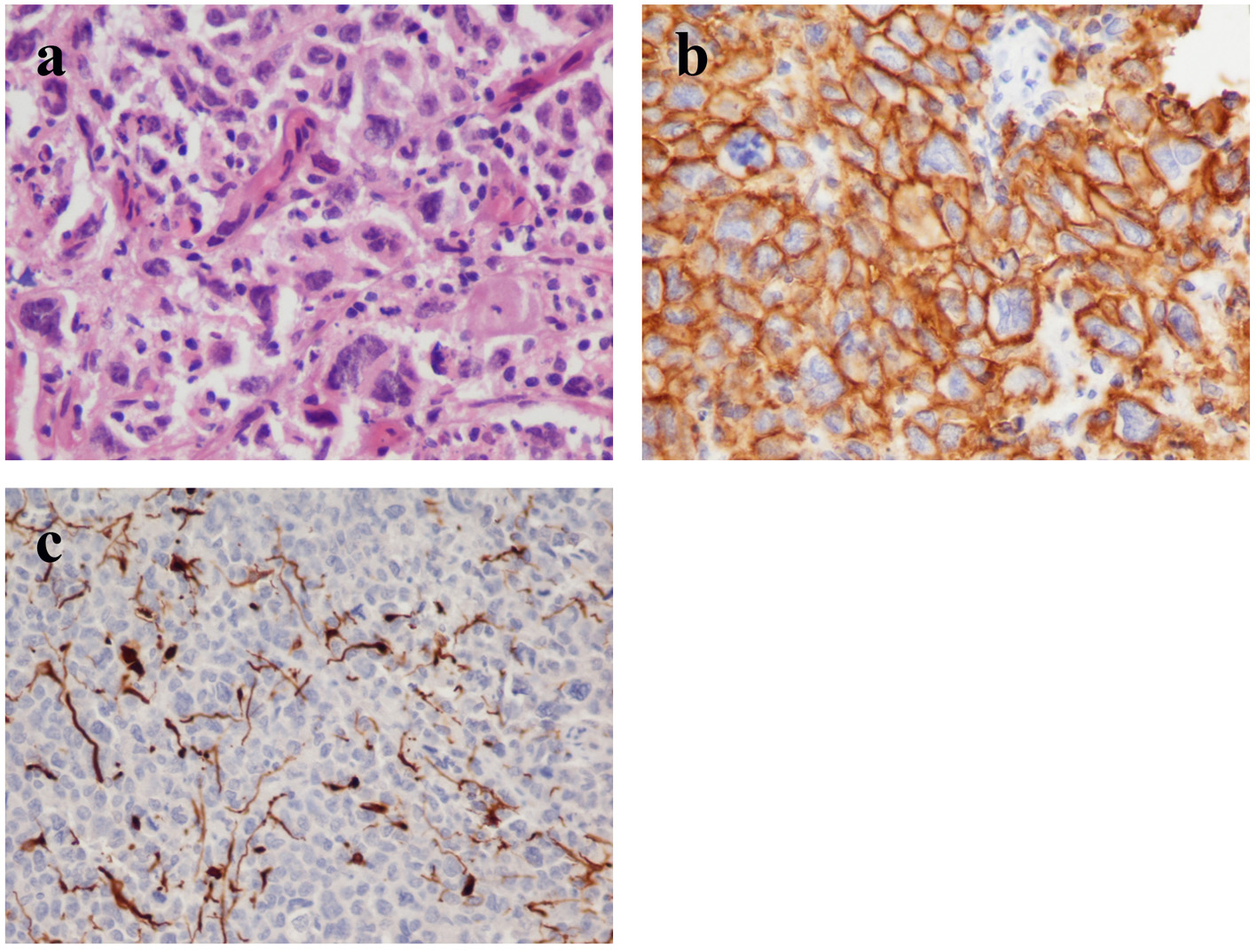
Figures