Journal of Clinical Medicine Research, ISSN 1918-3003 print, 1918-3011 online, Open Access
Article copyright, the authors; Journal compilation copyright, J Clin Med Res and Elmer Press Inc
Journal website http://www.jocmr.org

Case Report

Volume 7, Number 9, September 2015, pages 726-728


Benign Mature Teratoma in Anterior Mediastinum

Figures

Figure 1.
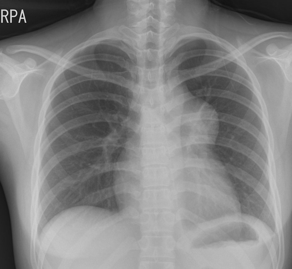
Figure 1. Chest X-ray showing protruding well-defined mass shadow in the left upper hilar space.
Figure 2.
Figure 2. Transthoracic echocardiography demonstrating a turbulent color Doppler signal in the pulmonary trunk. (A) Compressing mass in the pulmonary trunk in the parasternal short axis view (B) multilocular cystic mass (C). RA: right atrium; MPA: main pulmonary artery; AV: aortic valve.
Figure 3.
Figure 3. The chest CT showing a lobulated, inhomogeneous mass containing fat component in the mediastinum, compressing main pulmonary trunk (arrow), and no definite invasion to lung parenchyme (A, B, C).
Figure 4.
Figure 4. The pathologic findings. The excised specimen consisted of several fragments of heterogeneous soft tissue with disrupted outer surface (A). Photomicrographs of the mediastinal mass showed multiple areas of mature epidermis with sebaceous glands (B). In some areas of the mass, nests of respiratory epithelium and pancreatic tissue were observed (C). Foci of mature adipose tissue were also present in the tumor (D).