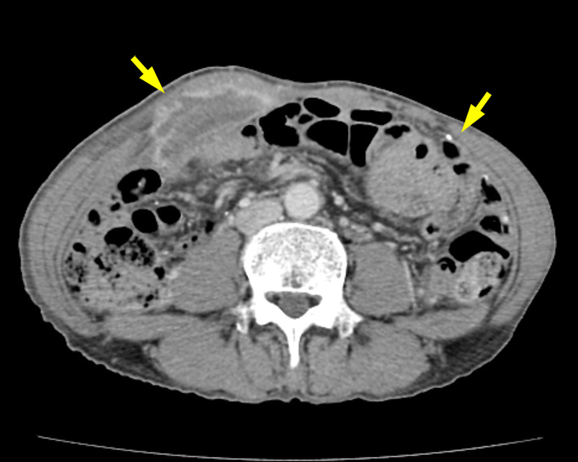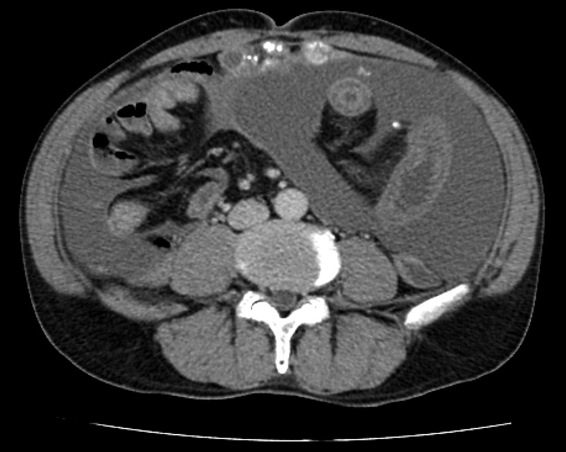
Figure 1. Abdominal computed tomographic scan before the initial operation showing massive ascites and peritoneal thickening with coarse calcifications.
| Journal of Clinical Medicine Research, ISSN 1918-3003 print, 1918-3011 online, Open Access |
| Article copyright, the authors; Journal compilation copyright, J Clin Med Res and Elmer Press Inc |
| Journal website http://www.jocmr.org |
Case Report
Volume 7, Number 7, July 2015, pages 571-574
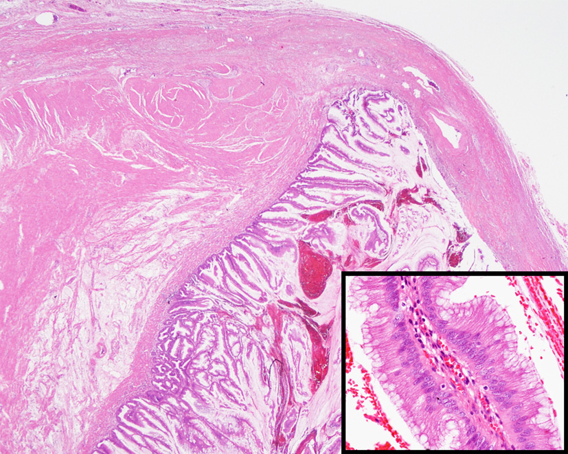
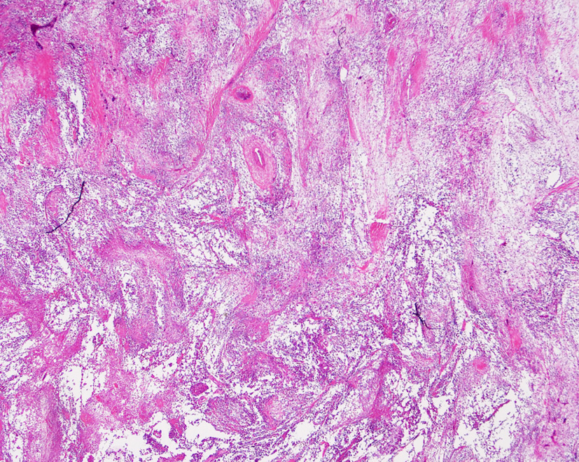
Transformation of Low-Grade Mucinous Neoplasm of the Appendix With Pseudomyxoma Peritonei to High-Grade Sarcomatoid Carcinoma
Figures