Journal of Clinical Medicine Research, ISSN 1918-3003 print, 1918-3011 online, Open Access
Article copyright, the authors; Journal compilation copyright, J Clin Med Res and Elmer Press Inc
Journal website http://www.jocmr.org

Case Report

Volume 7, Number 10, October 2015, pages 817-819


Isolated Right Ventricular Dilated Cardiomyopathy: An Early Diagnosis

Figures

Figure 1.
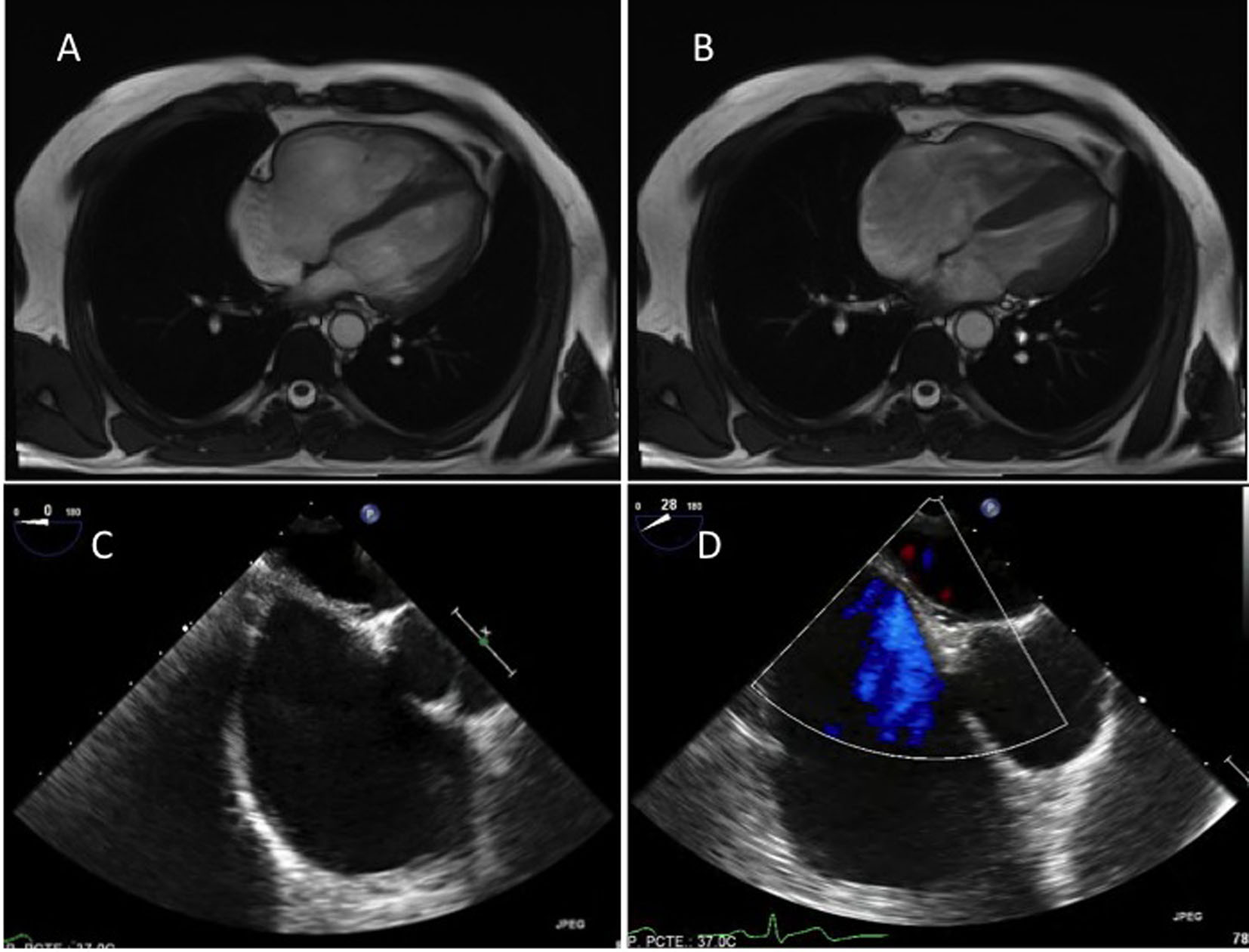
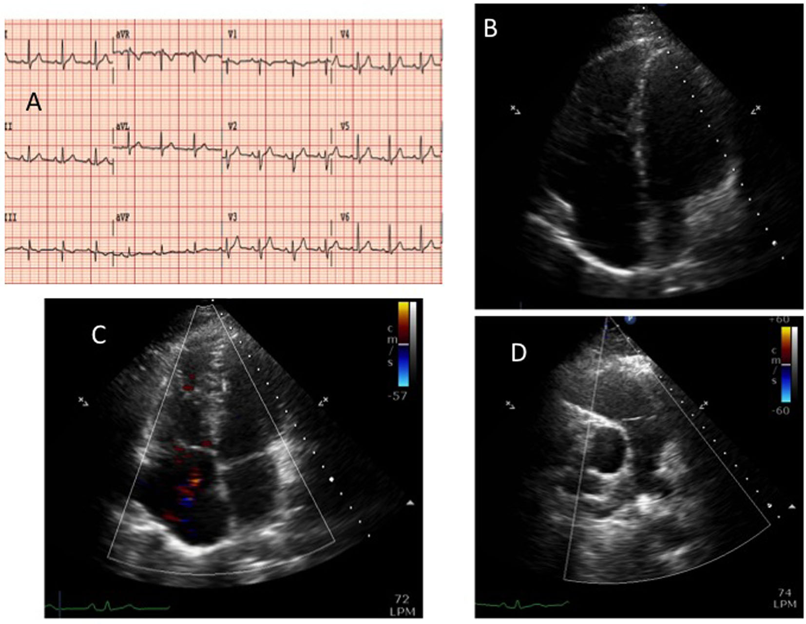
Figure 1. The 12-lead electrocardiogram (panel A). Transthoracic echocardiography: apical four-chamber view, diastolic frame (panel B) showing a severe dilatation of the right ventricle and color-Doppler systolic frame (panel C). Short axis parasternal view (panel D): dilatation of the right ventricle outflow tract but no dilatation in the pulmonary artery.
Figure 2.
Figure 2. Magnetic resonance, four-chamber view: diastolic (panel A) and systolic (panel B) frame, exhibiting normal right ventricular shortening. Transesophageal echocardiography, mid-esophageal plane (panel C) and also a color-Doppler (panel D) view exhibiting no interatrial shunt.