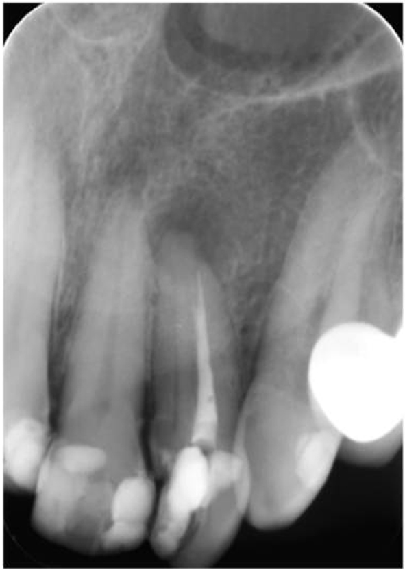
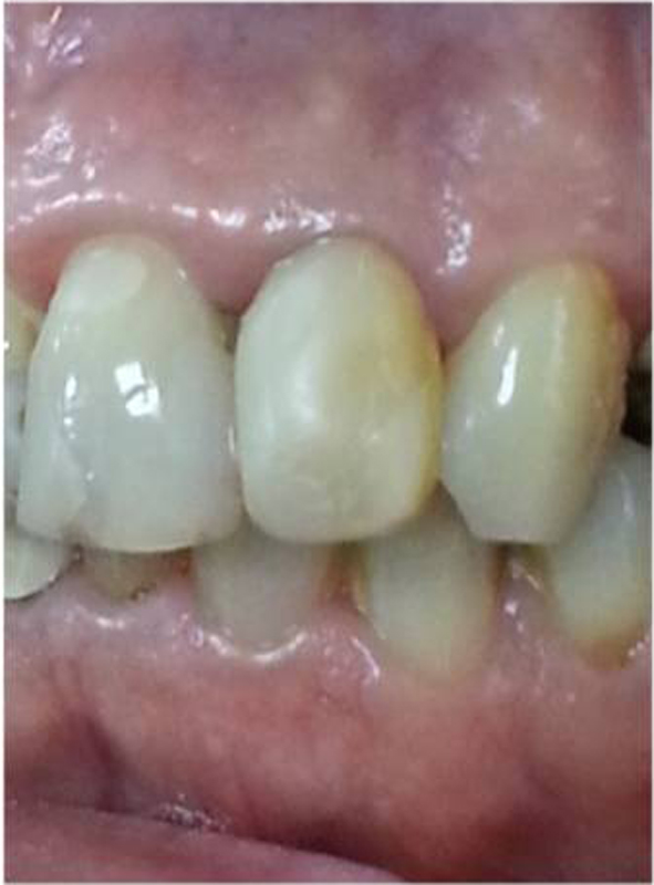
Figure 1. Preoperative periapical radiography.
| Journal of Clinical Medicine Research, ISSN 1918-3003 print, 1918-3011 online, Open Access |
| Article copyright, the authors; Journal compilation copyright, J Clin Med Res and Elmer Press Inc |
| Journal website http://www.jocmr.org |
Case Report
Volume 7, Number 7, July 2015, pages 560-563
Retreatment of a Maxillary Lateral Incisor With Two Separate Root Canals Confirmed With Cone Beam Computed Tomography
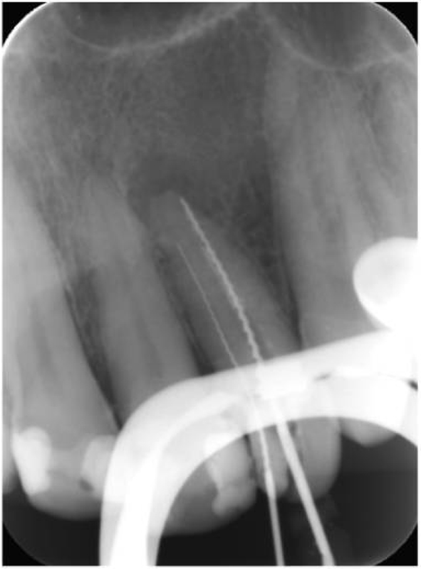
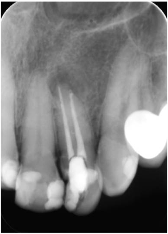
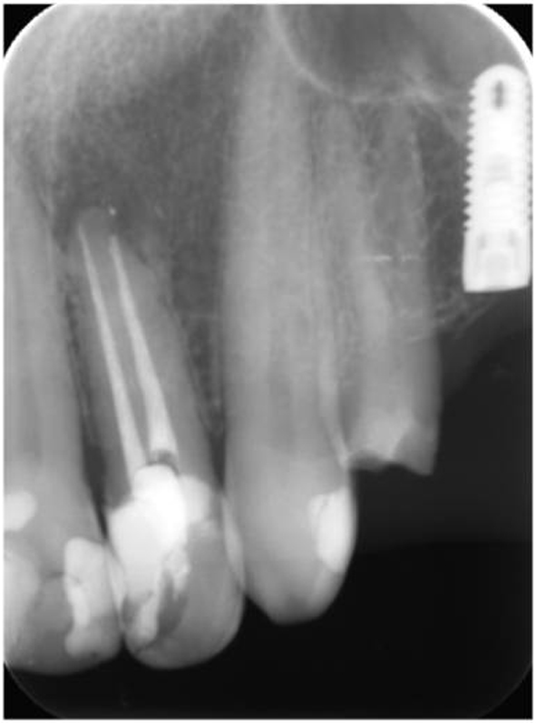
Figures

Table
| Category | Scores | Criteria |
|---|---|---|
| Retention | Alfa | No loss of restorative material |
| Charlie | Any loss of restorative material | |
| Color match | Alfa | Matches tooth |
| Bravo | Acceptable mismatch | |
| Charlie | Unacceptable mismatch | |
| Secondary caries | Alfa | No caries present |
| Charlie | Caries present | |
| Anatomic form | Alfa | Continuous |
| Bravo | Slight discontinuity, clinically acceptable | |
| Charlie | Discontinuous, failure | |
| Marginal adaptation | Alfa | Closely adapted, no detectable margin |
| Bravo | Detectable margin, clinically acceptable | |
| Charlie | Marginal crevice, clinical failure | |
| Surface texture | Alfa | Enamel-like surface |
| Bravo | Surface rougher than enamel, clinically acceptable | |
| Charlie | Surface unacceptable rough |