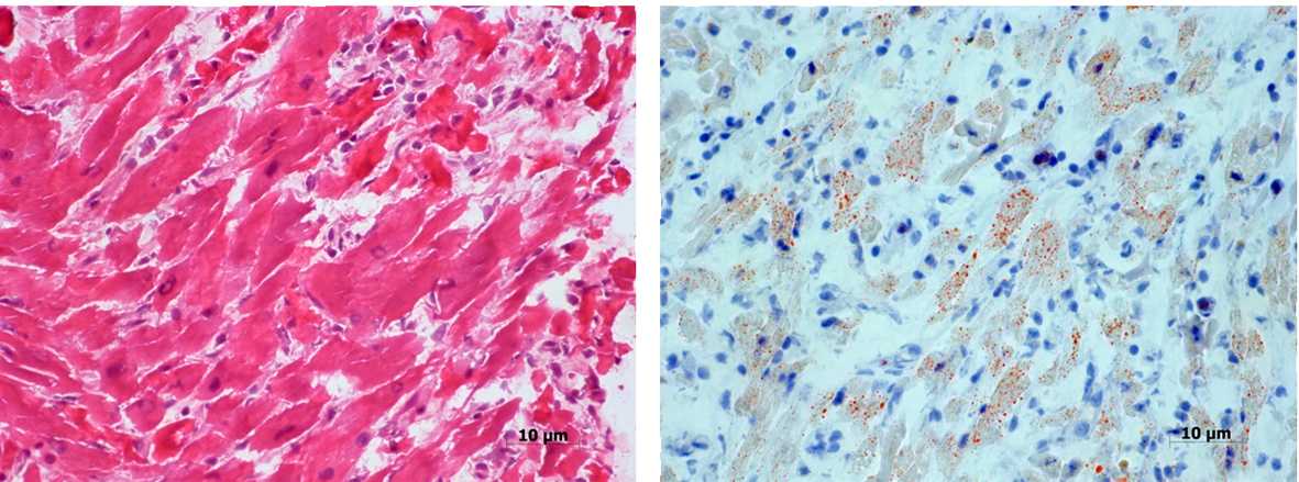
Figure 1. Section from the patient’s myocardium showing mononuclear inflammatory cell infiltration, degeneration and necrosis of some cardiac myocytes (A). Oil-Red-O stain reveals increased lipid in some fibers (B).
| Journal of Clinical Medicine Research, ISSN 1918-3003 print, 1918-3011 online, Open Access |
| Article copyright, the authors; Journal compilation copyright, J Clin Med Res and Elmer Press Inc |
| Journal website http://www.jocmr.org |
Original Article
Volume 7, Number 6, June 2015, pages 472-478
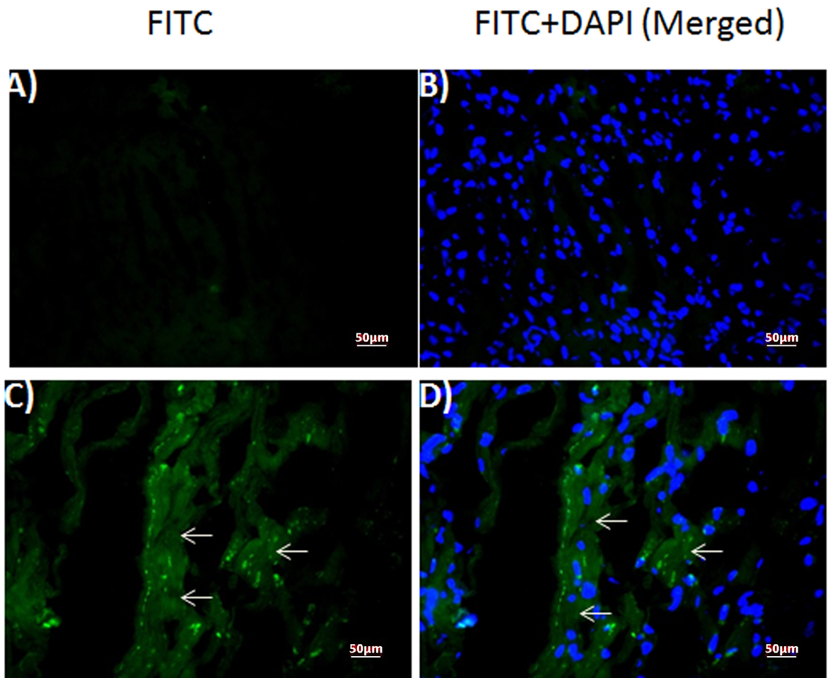
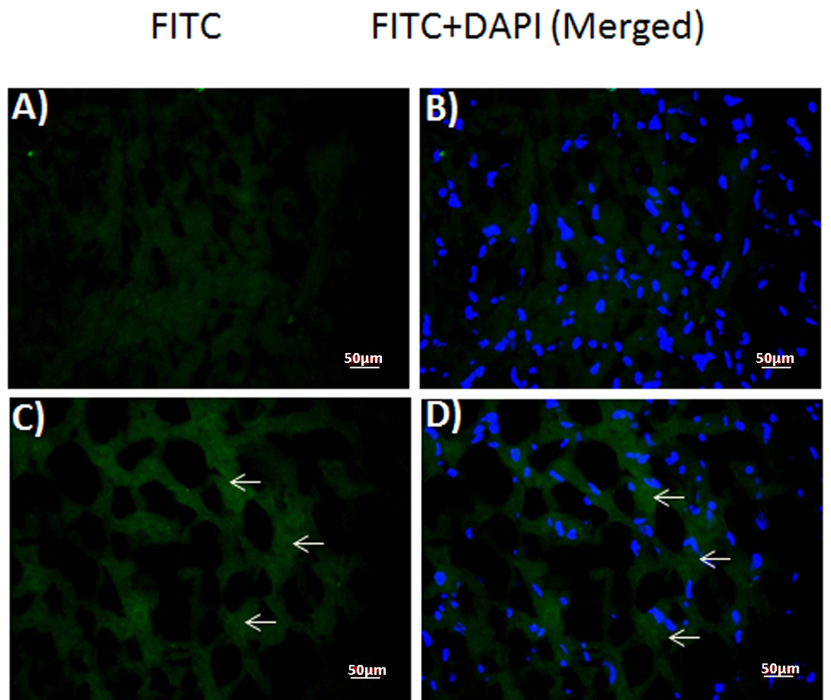
Determination of the Presence of Diphtheria Toxin in the Myocardial Tissue of Rabbits and a Female Subject by Using an Immunofluorescent Antibody Method
Figures