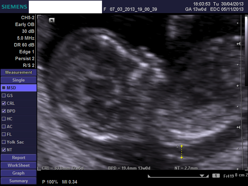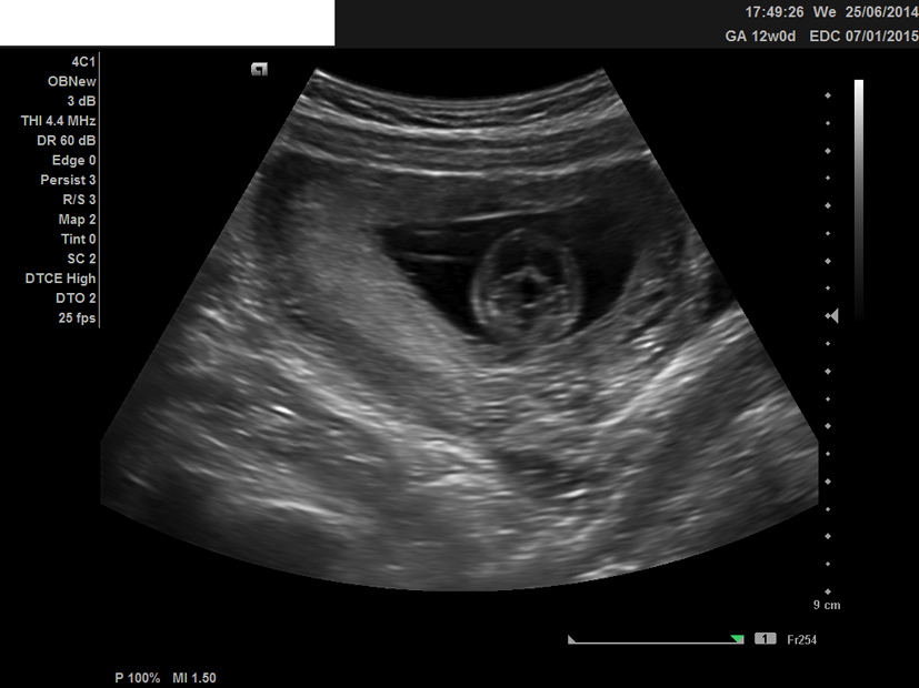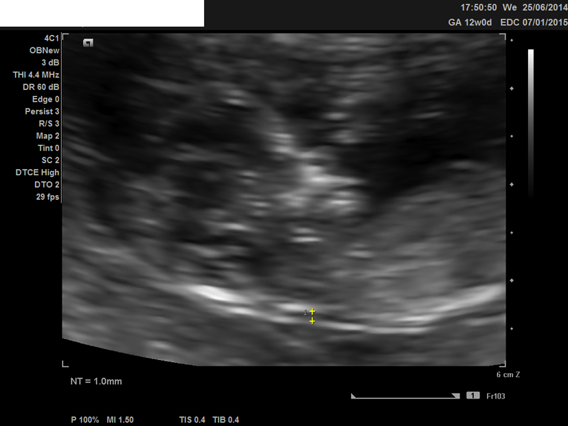
Figure 1. Ultrasound image of the second fetus at 12 weeks, showing the nuchal translucency and present nasal bone.
| Journal of Clinical Medicine Research, ISSN 1918-3003 print, 1918-3011 online, Open Access |
| Article copyright, the authors; Journal compilation copyright, J Clin Med Res and Elmer Press Inc |
| Journal website http://www.jocmr.org |
Case Report
Volume 7, Number 6, June 2015, pages 495-498
Variation of Ultrasound Findings in the First Trimester Examination of Recurrent Cases With Trisomy 21
Figures