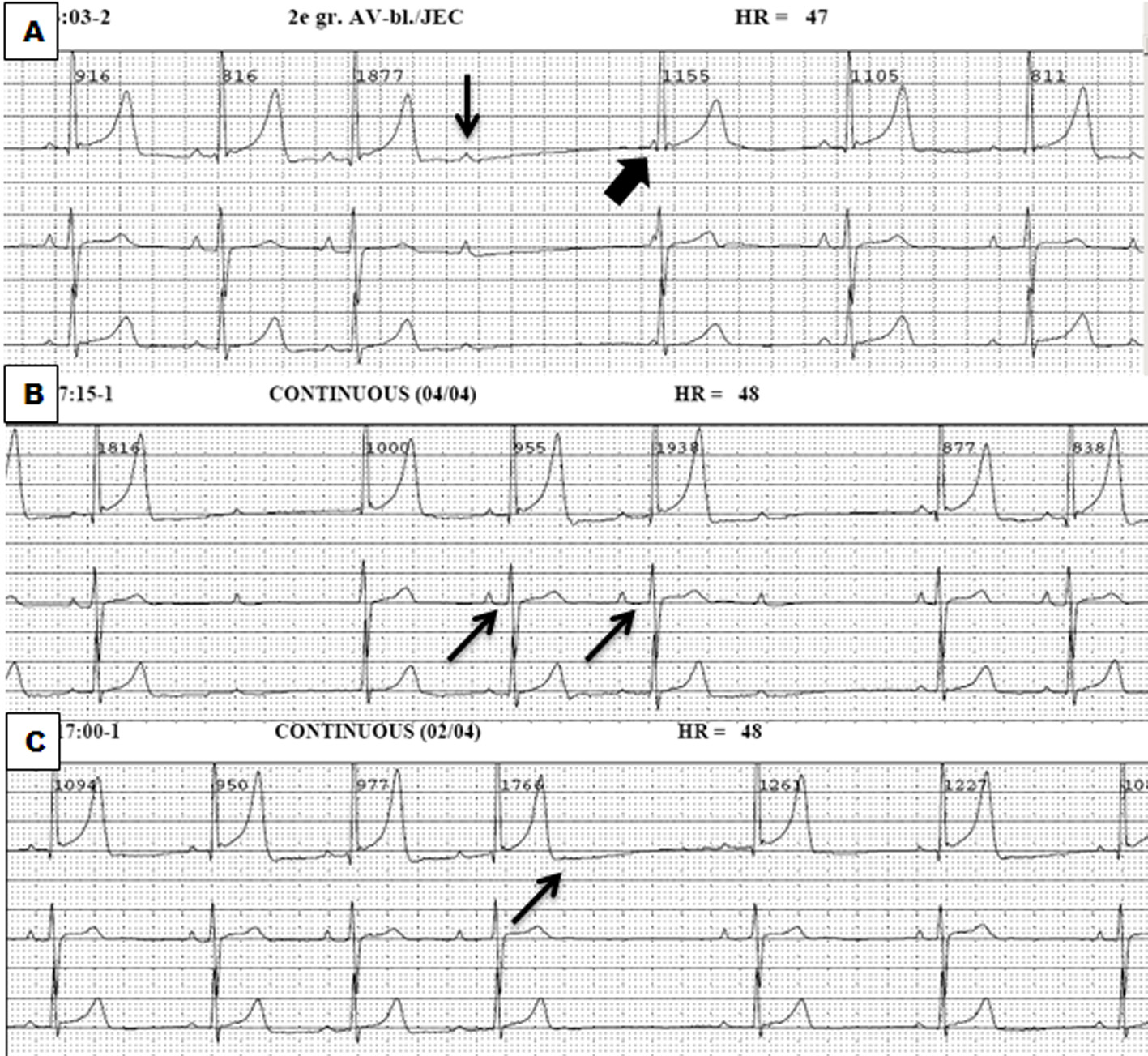
Figure 1. Holter recordings demonstrating a second degree atrioventricular block type II (thin arrow), sometimes followed by a junctional escape complex (thick arrow) (A), second degree atrioventricular block type I (B) and sinus arrests (C).
| Journal of Clinical Medicine Research, ISSN 1918-3003 print, 1918-3011 online, Open Access |
| Article copyright, the authors; Journal compilation copyright, J Clin Med Res and Elmer Press Inc |
| Journal website http://www.jocmr.org |
Case Report
Volume 7, Number 4, April 2015, pages 278-281
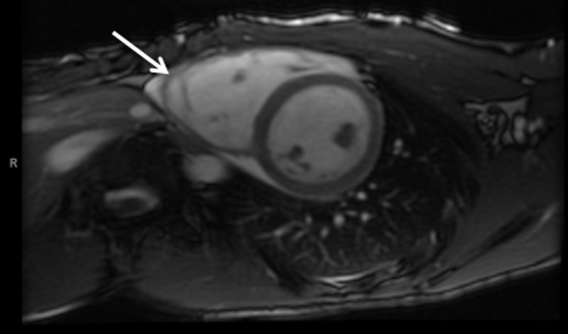
Bradyarrhythmias: First Presentation of Arrhythmogenic Right Ventricular Cardiomyopathy?
Figures