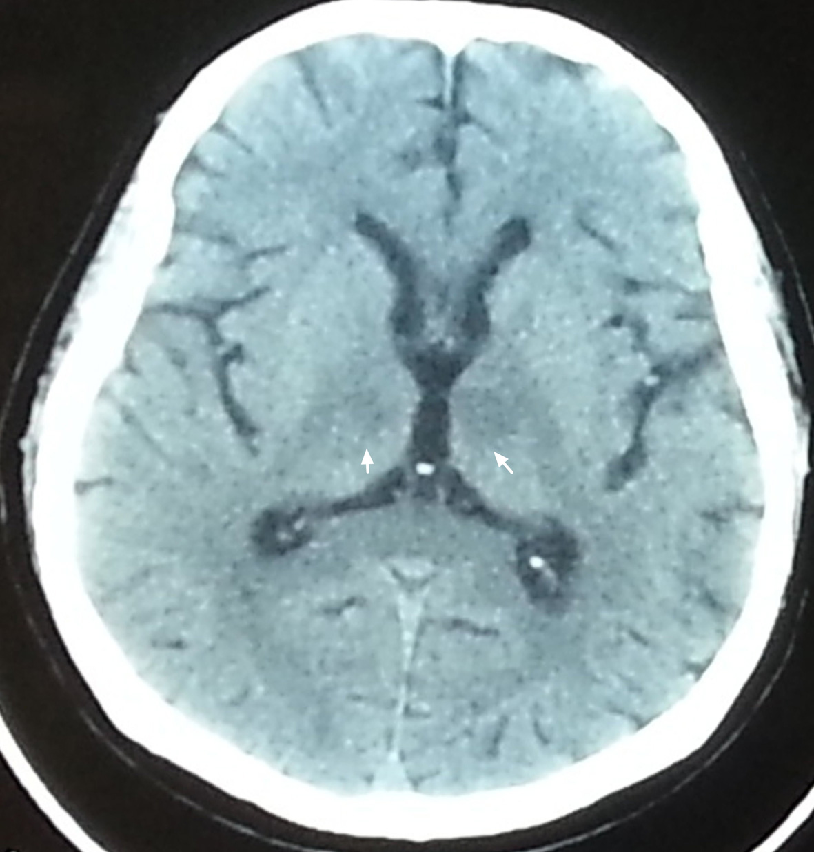
Figure 1. Axial non-contrast brain CT revealed faint hypodense lesion in bilateral paramedian thalamus (arrow).
| Journal of Clinical Medicine Research, ISSN 1918-3003 print, 1918-3011 online, Open Access |
| Article copyright, the authors; Journal compilation copyright, J Clin Med Res and Elmer Press Inc |
| Journal website http://www.jocmr.org |
Case Report
Volume 7, Number 2, February 2015, pages 126-128
Artery of Percheron Occlusion in an Elderly Male: A Case Report
Figures