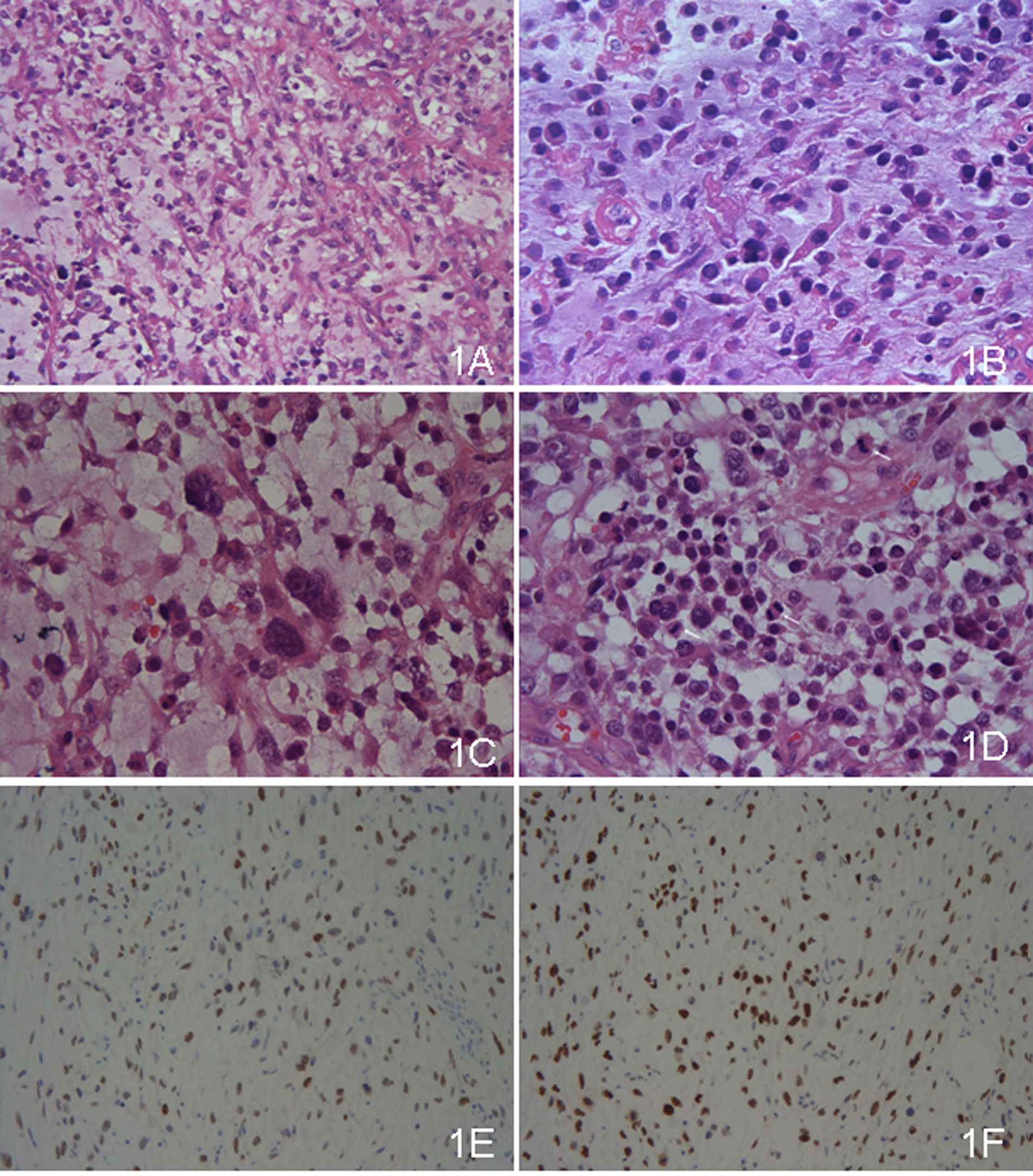
Figure 1. (A) The tumor was composed of abundant myxoid stroma comparted with fibrous tissues and veins, in which the tumor cells floated. Most of the tumor cells were medium, whereas some were large. The cells were lack of cohesion (×100, HE). (B) The tumor cells had ample amount of eosinophilic cytoplasms with coarse and prominent nuclei. The cytoplasms were irregular, some like the red streamer (×400, HE). (C) The giant odd cells were present (×400, HE). (D) Mitosis figures were common, and three were marked in the high power field (×400, HE). (E) WT-1 was positive. (F) Ki-67 labeling index was high.