Figures
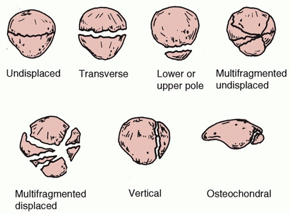
Figure 1. Descriptive classification of patellar fractures [5]. Reproduced with permission.
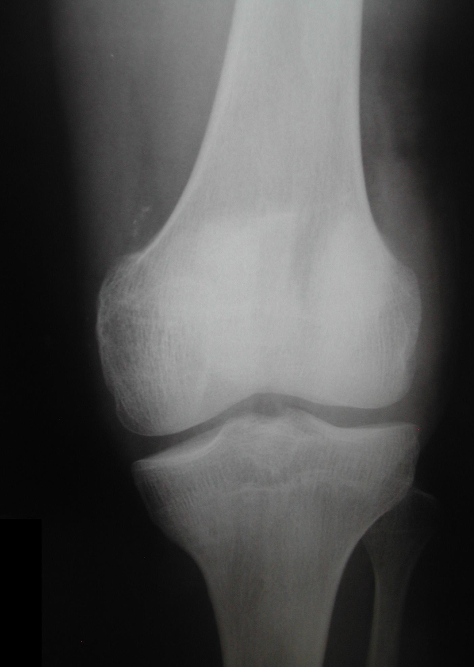
Figure 2. Anteroposterior radiograph demonstrated a vertical displaced patella fracture. Note that the deviated fragments are separated more than 3 mm.
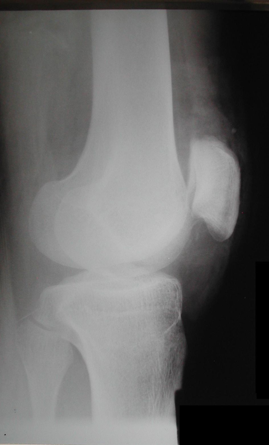
Figure 3. Lateral radiograph demonstrated the fracture by the articular gap in patella. Note the difference between the lateral and the medial side of patella caused by the displaced vertical trace of fracture.
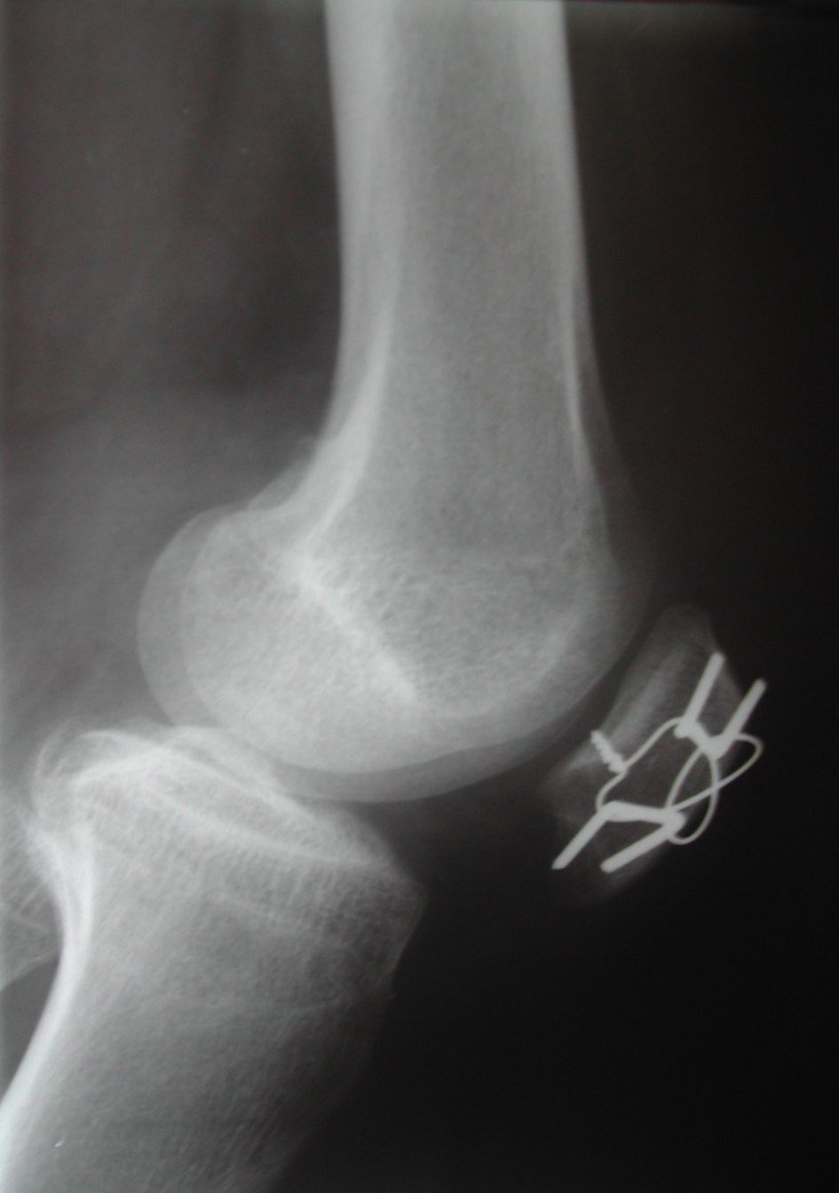
Figure 4. Seven days postoperatively, lateral radiograph demonstrated the total reduction of the patella fracture without joint gap and the use of the modified tension band.
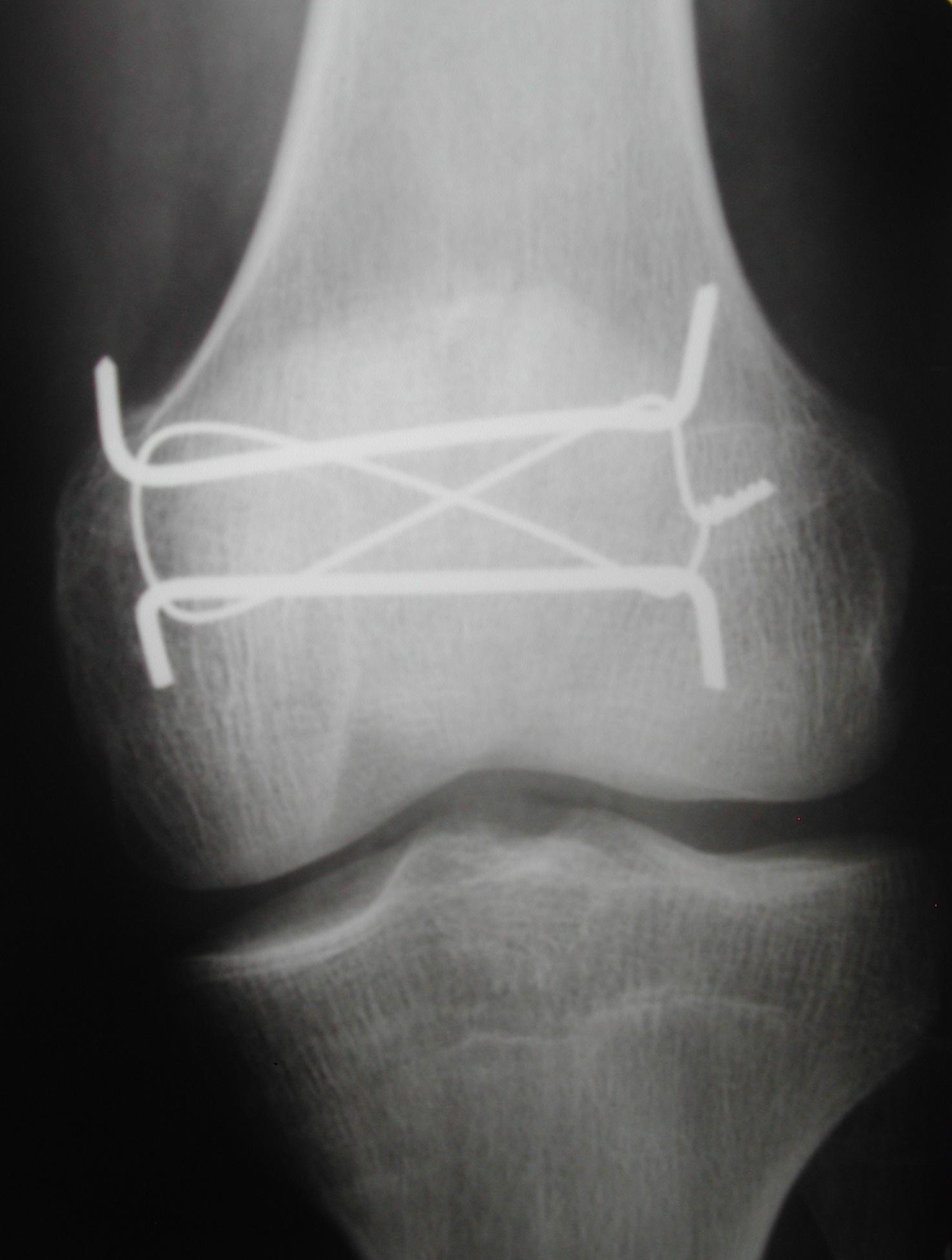
Figure 5. Seven days postoperatively, anteroposterior radiograph demonstrated the reduction of the separated fragments and the 3 mm distance between the fragments was gone. This incidence has shown better modified tension band.
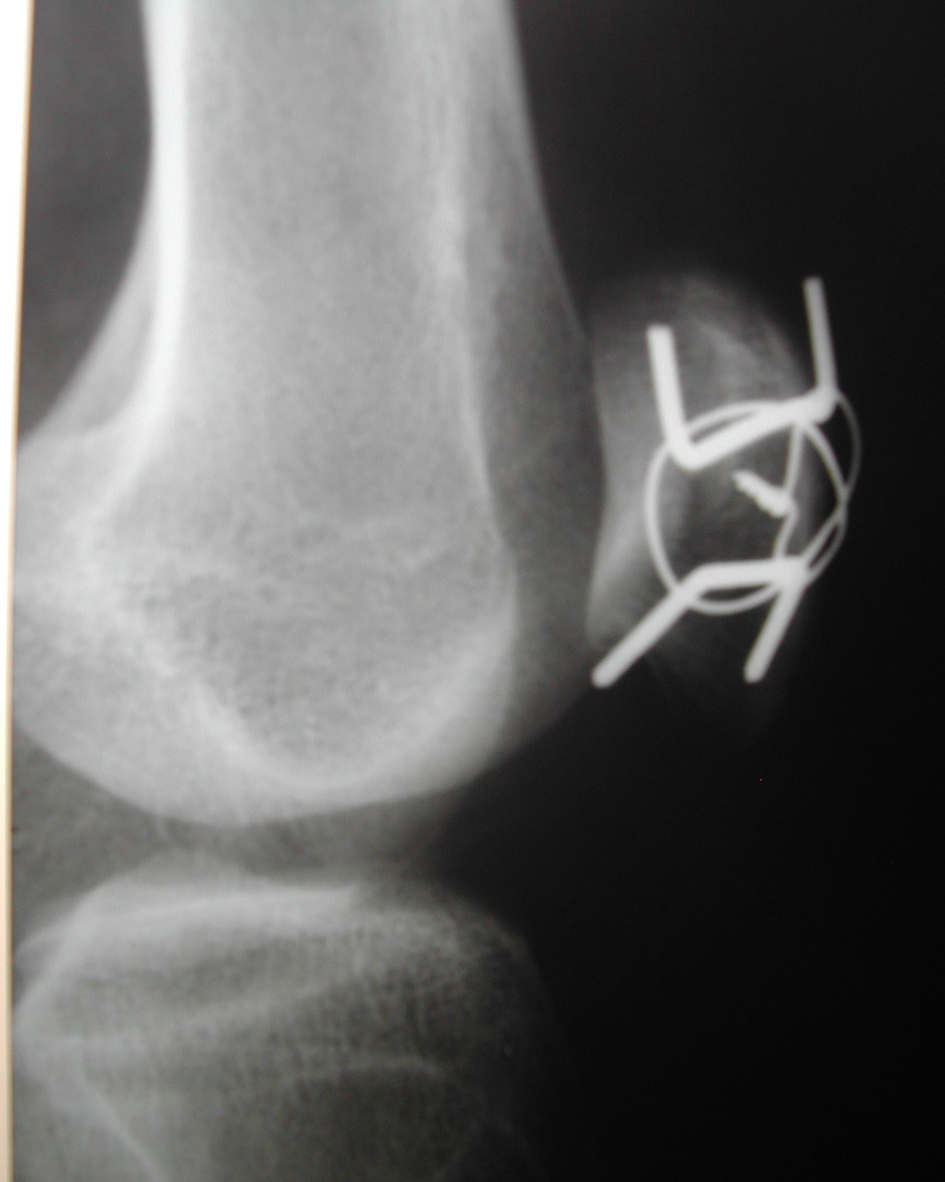
Figure 6. Seven days postoperatively, oblique radiograph demonstrated better articular surface without joint gaps.
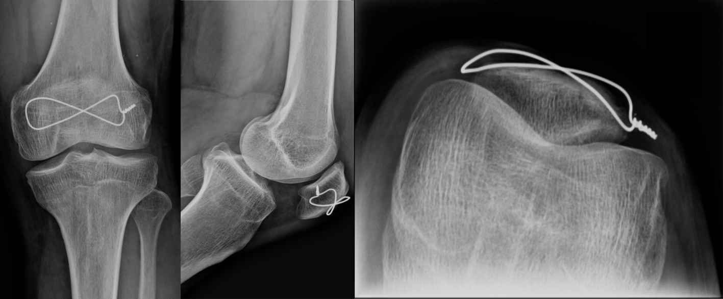
Figure 7. Twelve months postoperatively, no arthritis sign, the complete consolidation without articular gaps and the removal of the K-wires were shown. Anteroposterior radiograph (left side), lateral radiograph (center) and axial radiograph (right side).