Journal of Clinical Medicine Research, ISSN 1918-3003 print, 1918-3011 online, Open Access
Article copyright, the authors; Journal compilation copyright, J Clin Med Res and Elmer Press Inc
Journal website http://www.jocmr.org

Case Report

Volume 7, Number 2, February 2015, pages 122-125


Successful Surgical Treatment of Mature Teratoma Arising From the Sella

Figures

Figure 1.
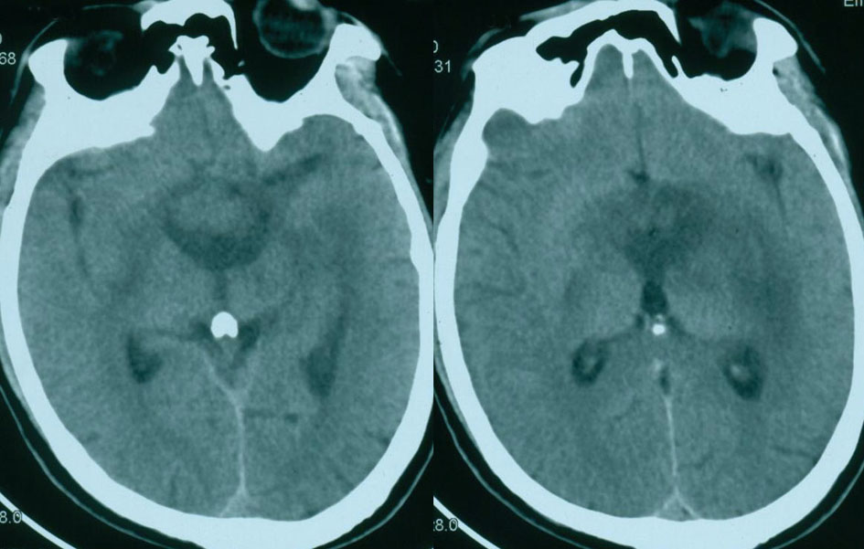
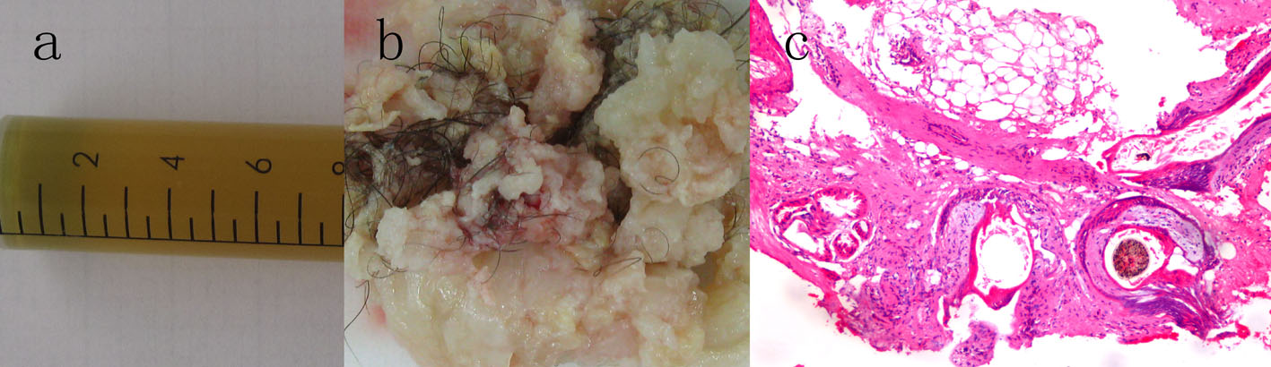
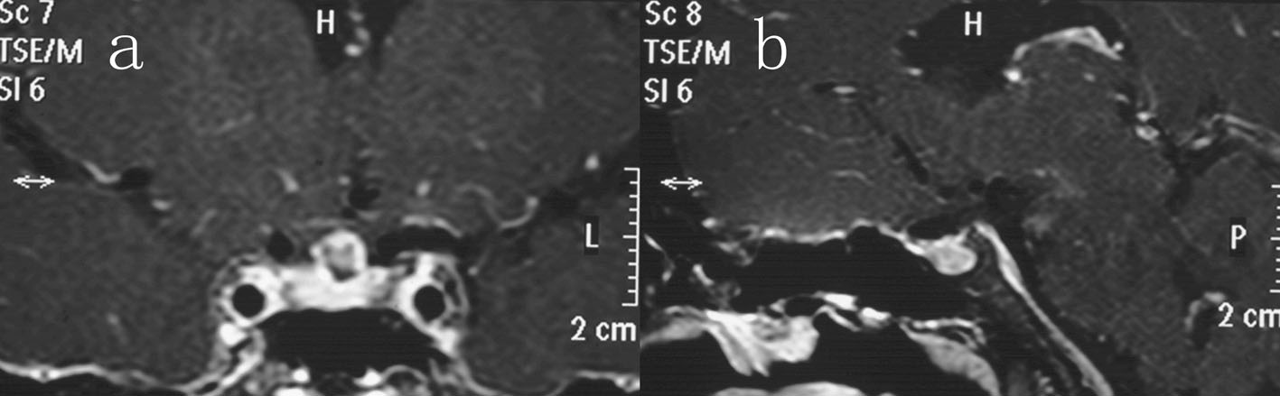
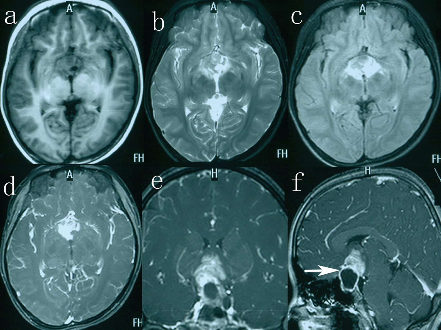
Figure 1. The solid part on axial T1-weighted imaging (a), T2-weighted imaging (b), fluid-attenuated inversion recovery image (c), axial (d) T1-weighted imaging with gadolinium. Coronal (e) and sagittal (f) image, enhanced with gadolinium, showing the tumor consisting of two components, an intrasellar cystic area and a suprasellar solid area (white arrow).
Figure 2.
Figure 2. CT showing a suprasellar mass with moderate dilation of the lateral ventricles.
Figure 3.
Figure 3. (a, b) Intraoperative photograph revealing the dark yellow cystic fluid and the solid tumor tissue. (c) Photomicrograph of the lesion demonstrating a mature teratoma with adipose tissue, hair, sebaceous glands, and stratified squamous epithelium. Hematoxylin and eosin stain, original magnifications × 100.
Figure 4.
Figure 4. Coronal (a) and sagittal (b) contrast-enhanced imaging after 12 months of follow-up showing complete excision of the sellar teratoma.