Journal of Clinical Medicine Research, ISSN 1918-3003 print, 1918-3011 online, Open Access
Article copyright, the authors; Journal compilation copyright, J Clin Med Res and Elmer Press Inc
Journal website http://www.jocmr.org

Case Report

Volume 7, Number 4, April 2015, pages 270-273


Atrial Flutter Ablation and Risk of Right Coronary Artery Injury

Figures

Figure 1.
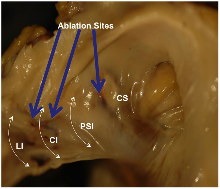
Figure 1. Gross examination of the right atrium shows white area of discoloration along the cavo-tricuspid isthmus region and an area of hemorrhage between the coronary sinus and the tricuspid valve. Three levels of the cavo-tricuspid isthmus are shown: lateral isthmus, central isthmus and paraseptal isthmus. A projected course of the right coronary artery (red line) demonstrates its anatomic relation to the cavo-tricuspid isthmus. CTI: cavo-tricuspid isthmus; FO: foramen ovale; TV: tricuspid valve; CS: coronary sinus; RCA: right coronary artery; TT: tendon of Todaro; LI: lateral isthmus; CI: central isthmus; PSI: paraseptal isthmus.
Figure 2.
Figure 2. Magnified right anterior oblique view of the internal structures of the right atrium. Ablation sites are shown (blue arrows). LI: lateral isthmus; CI: central isthmus; PSI: paraseptal isthmus; CS: coronary sinus.
Figure 3.
Figure 3. Magnified view of the distal right coronary artery shows brown discoloration of the epicardial fat.
Figure 4.
Figure 4. Microscopic examination shows cross-sections of the right coronary artery taken from the region of the ablation site demonstrating fat necrosis (red asterisk) and reactive changes to the fat and soft tissues of the right atrioventricular junction. Within the one-fifth of the vessel’s media, the smooth muscle cells have loss of nuclei (lack of blue colored nuclei) as shown by the black asterisk when compared to the normal smooth muscle cells with preserved nuclei (blue asterisk). Right coronary artery is narrowed by calcified atherosclerotic plaque with areas of acute plaque hemorrhage.