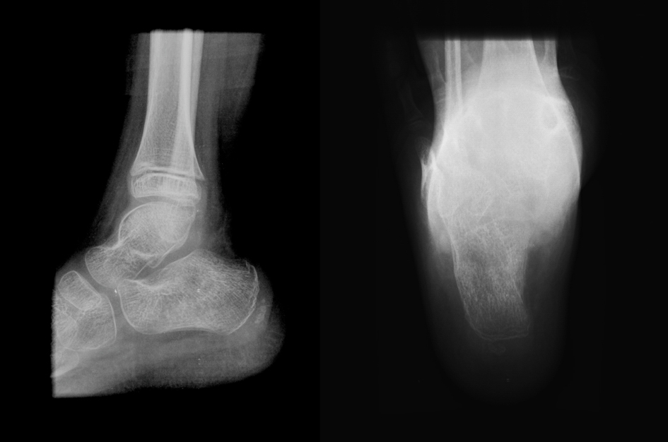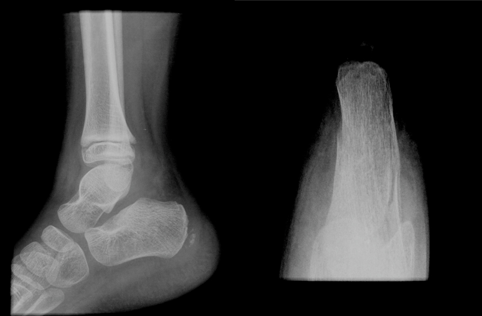
Figure 1. Lateral and axial image of the right calcaneal. Lateral view has not shown signs of fracture. The axial view revealed a calcaneal body fracture. Both images do not clarify if the fracture affected the joint.
| Journal of Clinical Medicine Research, ISSN 1918-3003 print, 1918-3011 online, Open Access |
| Article copyright, the authors; Journal compilation copyright, J Clin Med Res and Elmer Press Inc |
| Journal website http://www.jocmr.org |
Case Report
Volume 7, Number 1, January 2015, pages 52-55
An Atypical Calcaneal Fracture in a Child: A Literature Review Concerning the Treatment
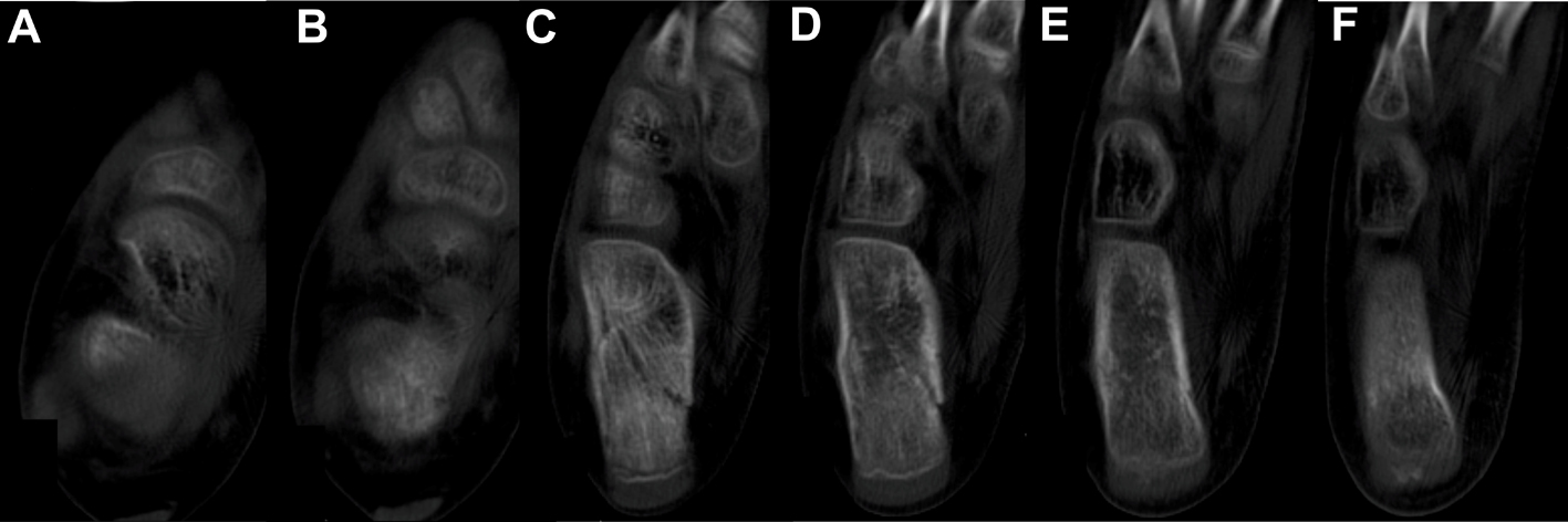
Figures