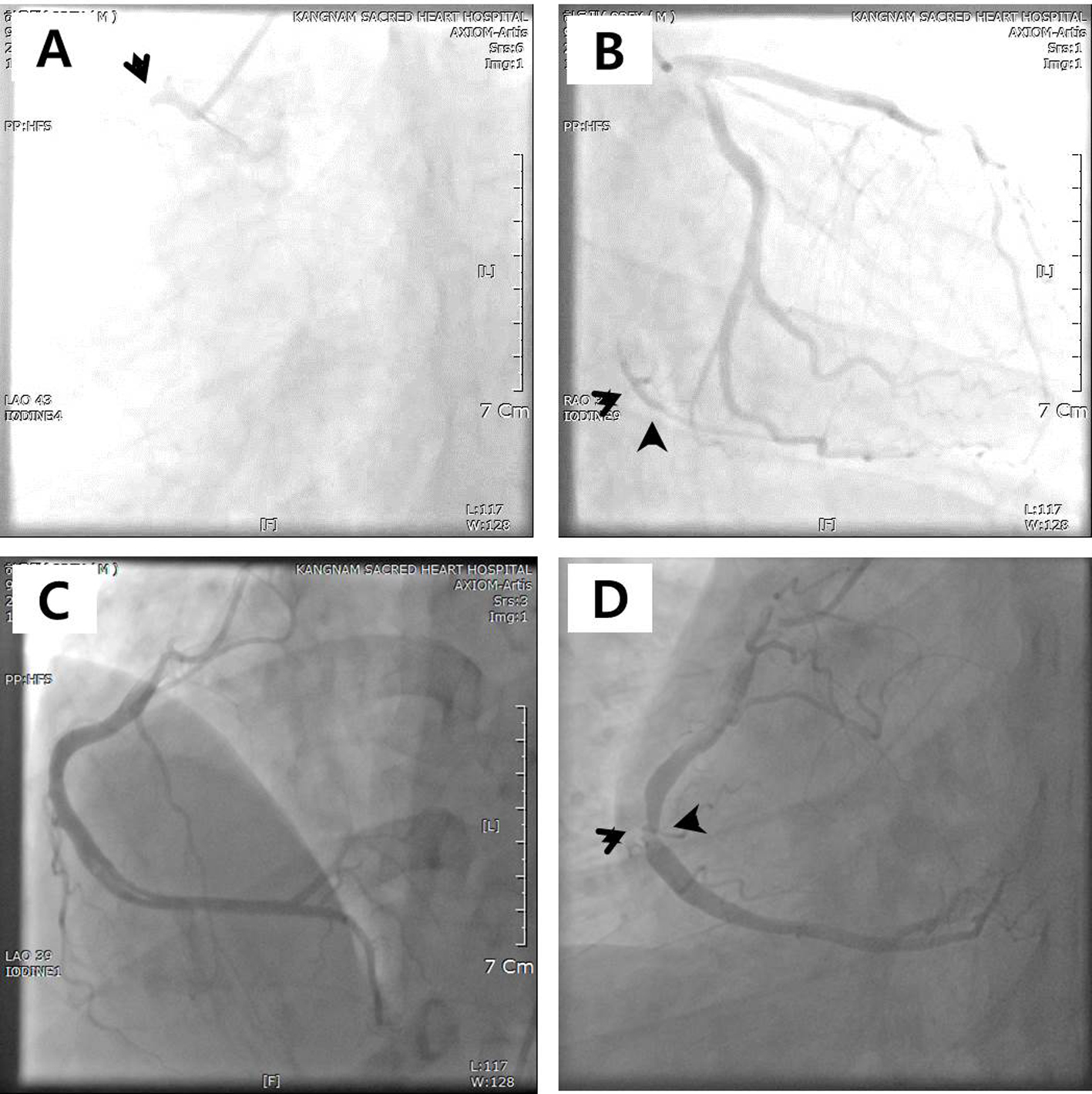
Figure 1. (A, B) Coronary computed tomography showing normal coronary arteries without stenosis.
| Journal of Clinical Medicine Research, ISSN 1918-3003 print, 1918-3011 online, Open Access |
| Article copyright, the authors; Journal compilation copyright, J Clin Med Res and Elmer Press Inc |
| Journal website http://www.jocmr.org |
Case Report
Volume 7, Number 1, January 2015, pages 62-64
Young Patient Presenting Acute Coronary Syndrome
Figures