Figures
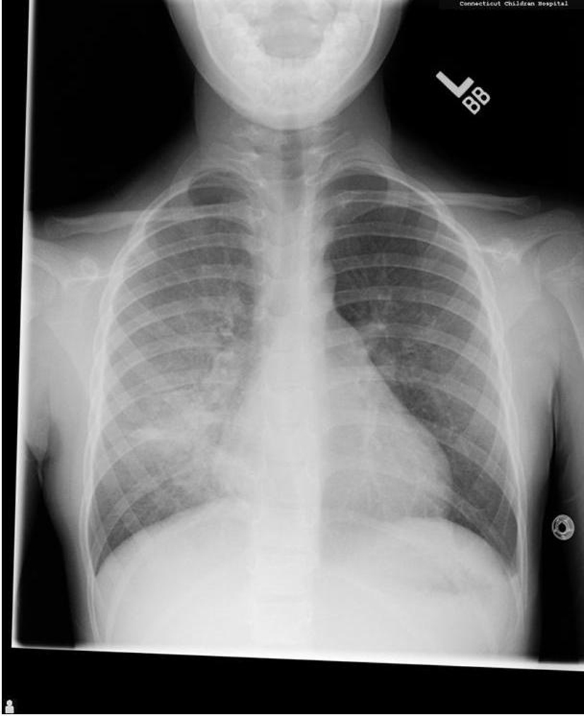
Figure 1. Initial chest X-ray demonstrated a diffuse airspace filling process throughout the right lung and a small airspace opacity in the left upper lobe.
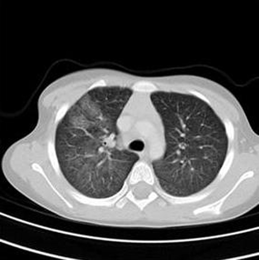
Figure 2. Initial chest CT demonstrated the extent of the airspace filling process. Ground glass opacities were seen in all lobes of the right lung with ground glass opacity identified in the left upper and left lower lobes to a lesser extent. In addition, there is dense right middle lobe consolidation.
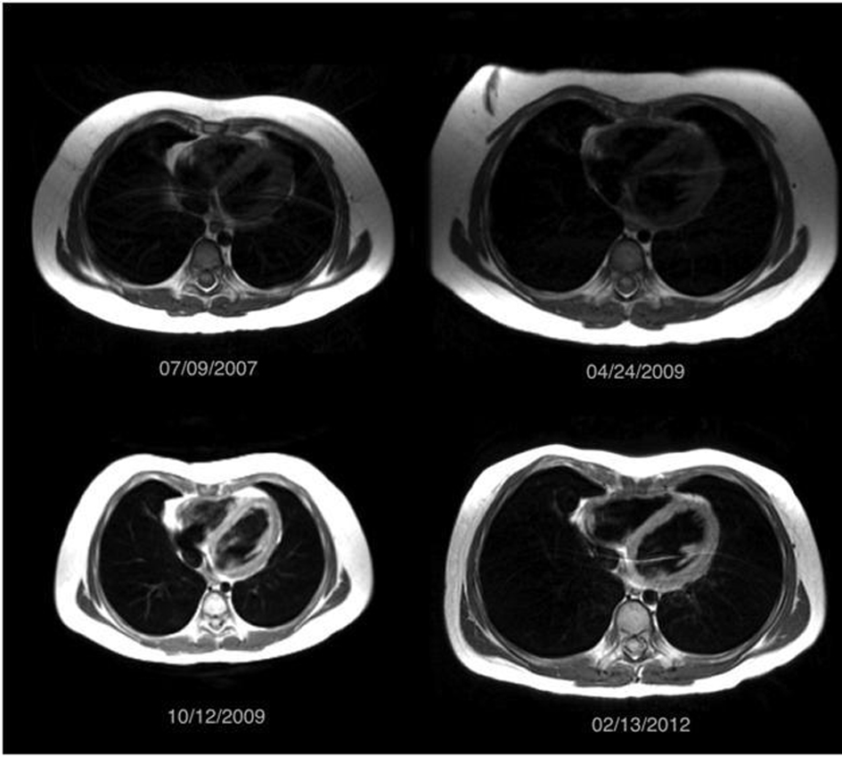
Figure 3. Multiple surveillance MRI scans obtained between July 2007 and February 2012 demonstrated clear lungs without evidence of an abnormal airspace filling process. The patient was asymptomatic during this time period.
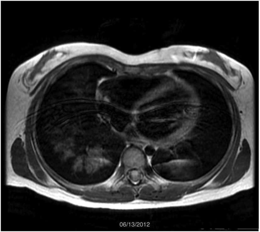
Figure 4. MRI scan obtained on June 2012 demonstrated diffuse airspace opacities in the right lower lobe and left lower lobe. Clinically, the patient was experiencing hemoptysis and found to be in an exacerbation of her condition.
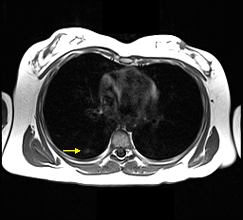
Figure 5. MRI scan obtained November 2012. Axial T1 image showed multiple small peripheral airspace opacities in the right lower lobe (yellow arrow), which was compatible with alveolar hemorrhage given the patient’s history and clinical presentation.