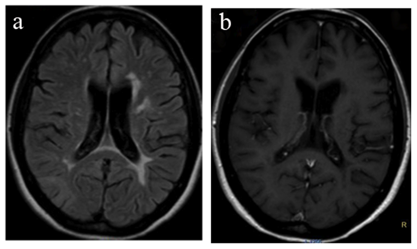
Figure 1. MRI on initial relapse reveals a new lesion in the subcortical white matter of the right parietal lobe with high signal on the FLAIR sequence (arrows, a) and mild contrast enhancement (arrow, b).
| Journal of Clinical Medicine Research, ISSN 1918-3003 print, 1918-3011 online, Open Access |
| Article copyright, the authors; Journal compilation copyright, J Clin Med Res and Elmer Press Inc |
| Journal website http://www.jocmr.org |
Case Report
Volume 7, Number 1, January 2015, pages 65-68
Immune Reconstitution Inflammatory Syndrome Mimicking Progressive Multifocal Leucoencephalopathy in a Multiple Sclerosis Patient Treated With Natalizumab: A Case Report and Review of the Literature
Figures