


| Journal of Clinical Medicine Research, ISSN 1918-3003 print, 1918-3011 online, Open Access |
| Article copyright, the authors; Journal compilation copyright, J Clin Med Res and Elmer Press Inc |
| Journal website http://www.jocmr.org |
Short Communication
Volume 1, Number 2, June 2009, pages 121-122
An Extraosseous Plasmacytoma of the Nasopharynx
Ravinder Singh Natta, b, Gerry O’Sullivana
aWirral University Teaching Hospitals NHS Trust, Arrowe Park Road, Merseyside CH49 5PE, UK
bCorresponding author: Wirral University Teaching Hospitals NHS Trust, Department of Otolaryngology, Arrowe Park Road, Merseyside CH49 5PE, UK
Manuscript accepted for publication May 27, 2009
Short title: Plasmacytoma of Nasopharynx
doi: https://doi.org/10.4021/jocmr2009.05.1240
| Abstract | ▴Top |
A 75-year-old long-term male smoker and poorly controlled hypertensive presented with a 3-month history of intermittent epistaxis refractory to cauterisation with silver nitrate. Nasopharyngeal examination revealed a mass in the post nasal space. An urgent endoscopic biopsy confirmed an extraosseous plasmacytoma. Post operative radiotherapy was scheduled. No evidence of recurrence of disease following completion of treatment has been detected during clinical surveillance.
Keywords: Epistaxis; Nasopharynx; Biopsy; Plasmacytoma; Radiotherapy
| ▴Top |
A 75-year-old long-term male smoker and poorly controlled hypertensive presented with a 3-month history of intermittent epistaxis refractory to cauterisation with silver nitrate. Nasopharyngeal examination revealed a smooth mass extending inferiorly from the right fossa of Rossenmuller and effacing the posterior pharyngeal wall (Fig 1). An urgent endoscopic examination and excisional biopsy was scheduled. Haematoxylin and eosin staining confirmed a dense infiltration of plasmacytoid cells. Immunocytochemistry confirmed malignant plasma cells consistent with an extraosseous plasmacytoma.
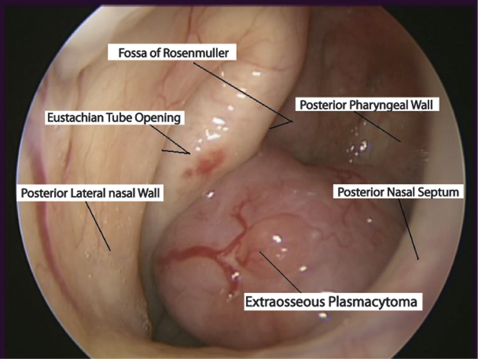 Click for large image | Figure 1. Location of the extraosseous plasmacytoma |
Serum and urine assays for Bence Jones protein were negative and there was no evidence of gammopathy. A bone marrow biopsy and Computed Tomography and Magnetic Resonance imaging did not demonstrate any metastasis or skeletal involvement. The Excisional biopsy had been complete and the patient underwent radical radiotherapy with a 45Gy dose in 20 fractions of the nasopharyngeal field.
No evidence of recurrence of disease following completion of treatment has been detected during clinical surveillance.
A plasmacytoma is a very rare discrete solitary mass of neoplastic monoclonal plasma cells, first described by Schridde in 1905 [1]. They are classified into one of two categories; soft tissue and skeletal origin. Extramedullary plasmacytomas represent 3% of plasma cell neoplasms and commonly (80%) originate in the head and neck region [2]. They represent approximately 4% of nasal cavity tumours. There is a greater male preponderance and they occur during the fifth and seventh decades of life [3]. The aetiology remains unknown but viral pathogenesis and chronic irritation from inhaled irritants have been suggested [4].
Tissue biopsy, serum electrophoresis (to exclude myeloma) and radiological skeletal survey with bone marrow biopsy to determine skeletal involvement is necessary for diagnosis. Treatment includes a combination of surgical excision and radiotherapy. Follow-up radiological and electrophoresis assessment is required after treatment to detect recurrences and progression to myeloma (10-30% frequency). The overall 10 year survival is 70% [5].
Acknowledgments
There are no competing interests to declare.
| References | ▴Top |
- Schridde H. Weitere Untersuchungen uber die Kornelungen der Plasmazellen. Centralbl Allg Pathol Pathol Anat. 1905;16:433-435.
- Paris J, Dessi P, Moulin G, Chrestian MA, Braccini F, Zanaret M. [Extramedullary plasmocytoma of the nasal cavity: a case report]. Rev Laryngol Otol Rhinol. (Bord)1999;120(5):343-345.
pubmed - Susnerwala SS, Shanks JH, Banerjee SS, Scarffe JH, Farrington WT, Slevin NJ. Extramedullary plasmacytoma of the head and neck region: clinicopathological correlation in 25 cases. Br J Cancer. 1997;75(6):921-927.
pubmed - Lomeo PE, McDonald JE, Finneman J, Shoreline . Extramedullary plasmacytoma of the nasal sinus cavities. Am J Otolaryngol. 2007;28(1):50-51.
pubmed - Straetmans J, Stokroos R. Extramedullary plasmacytomas in the head and neck region. Eur Arch Otorhinolaryngol. 2008;265(11):1417-1423.
pubmed
This is an open-access article distributed under the terms of the Creative Commons Attribution License, which permits unrestricted use, distribution, and reproduction in any medium, provided the original work is properly cited.
Journal of Clinical Medicine Research is published by Elmer Press Inc.